SAN FRANCISCO - The future of CT development cannot be known precisely, according to Dr. Willi Kalender, Ph.D., from the Medical Physics Institute of the University of Erlangen-Nuernburg in Germany. But it is sure to be bright, he said.
Kalender discussed the future of CT imaging at Wednesday's opening of the 2003 International Symposium on Multidetector-row CT, hosted by Stanford University. He warned that predicting the future of the dynamic technology was a guessing game, and that it would someday be easy to prove him wrong.
Still, having developed spiral CT in 1989 and advanced it markedly to the present day, Kalender is thought to be in a good position to guess. His first prediction was that the so-called slice race would continue.
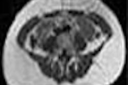
"There is no problem with detector technology (going) to a higher number of slices," Kalender said. "There is already a 256-row detector ... in early investigation. What is more of a problem is image reconstruction."
Several different detector designs that are already available -- including isotropic, non-isotropic, and adaptive arrays -- work very well, he said. The main limitation in coverage is the number of detector rows, which determines the speed of volume coverage.
In turn, the number of detector rows that can be acquired simultaneously is subject to some complications regarding the electronics needed to deliver all of the detector signals, as well as data transfer and storage quandaries. But these issues can be solved, he said. Kalender sees the need for new and more complex algorithms as the main limitation to increasing the number of detector rows.
The need for these algorithms results from the so-called cone beam problem. That is, with single-detector scanners, the beam is always in the center and encounters the same data at any angle. However, as the number of detectors increases, the data collected for each detector's data acquisition corresponds to the volume between two cones, rather than a flat plane. This leads to image distortion that is most pronounced for the outer cones.
As the number of detector rows increases, "we can't do as we have always done and take the (detector) row number and then take the orientation to construct the x-y plane for a given z position," he said.
Beyond four detectors, image distortion becomes pronounced, and formulae such as the advanced single-slice rebinning (ASSR) algorithm are needed to correct for the divergent beam angles. ASSR, developed by Kalender and colleagues, performs image reconstruction for arbitrarily oblique planes, which are fitted optimally to the spiral path.
This solution works well for the new 16-slice scanners, with very little image distortion, Kalender said. However, new solutions will be needed to handle greater numbers of detectors. Such algorithms are already in the works for up to 128 detectors, while for now the use of 256 detectors can only be simulated, he said.
Regarding radiation protection, Kalender said one of the main challenges was to communicate to the public that CT doses are already at a very low level. The United Nations International Atomic Energy Agency (IAEA) and other international organizations consider exposure under 200 mSv to be within the low-dose range, and nearly all CT scanning, even in multiple exposures, falls well below that level.
Dose information on scanners will become more sophisticated over the next several years, and along with automatic exposure controls will go a long way toward allaying patients' fears, he said. Dose-measurement technology similar to that used in planning radiation therapy already exists, he said, and will be applied to CT scanners.
"In the future, we will have to communicate dose to show we know what we're doing," Kalender said.
Other emerging technologies will also help minimize radiation exposures, he said, including automated tube current modulation, and a variety of noise reduction filters that improve noise behavior in reconstruction and enable lower-dose acquisitions.
"What we're shooting for has to be the combination of all those measures into automated exposure control," he said. "Automated exposure control has to guarantee that for a given image-quality level, we as radiologists shall not fiddle around with mAs or slice thickness. We just have to tell the machine, for example, 'I want a (20-second scan) isotropically with a noise level of 10 HU' -- and the rest has to be left to the algorithm."
Cardiac imaging will benefit significantly from more detector rows by detecting smaller calcifications earlier. ECG gating will fall by the wayside as cardiac motion is detected in the raw image data and subsequently corrected by algorithms, he said.
Many believe that flat-panel detectors are the future of CT, Kalender said. But while flat-panel detectors are improving rapidly and will be able to produce high spatial resolution, good low-contrast resolution will remain a challenge, and will be difficult to improve significantly. As a result, Kalender said he believes the use of area detector CT will be limited to special applications.
By Eric BarnesAuntMinnie.com staff writer
June 27, 2003
Related Reading
Radiotherapy access must be improved in developing world: UN, June 27, 2003
Multislice CT reveals more coronary segments than electron beam CT for CAD, May 22, 2003
Standard, low-dose CT calcium screening yield equivalent results, March 10, 2003
Flat-panel unit beats CR for digital mammography, March 7, 2003
Copyright © 2003 AuntMinnie.com