BOSTON - If it is to succeed, CT lung screening has a big job to do. The challenge -- longer survival through early detection and treatment -- can seem like a tall order for a disease that is responsible for 28% of all cancer deaths in the U.S., or 157,000 deaths per year for every 165,000 diagnosed. Moreover, three decades of lung screening trials have failed to increase survival. Largely for that reason, no U.S. healthcare organization recommends CT lung screening.
On the other hand, a revolution is underway in CT technology. Multidetector-row scanners and better software keep improving radiologists' ability to see lung abnormalities. As for the trials, they have brought huge amounts of information to light about lung cancer, and the benefits of early detection. Lung cancer patients are known to have a better chance of being cured when the disease is found through screening rather than clinical presentation.
At a conference organized by the Society of Computed Body Tomography and Magnetic Resonance, titled, "Medically Principled CT Screening," presenters shared techniques, theories, and controversies surrounding CT screening and related technologies.
In her talk, Dr. Ella Kazerooni, director of thoracic radiology at the University of Michigan in Ann Arbor, summed up the evidence in favor of -- and against -- the use of regular CT lung exams.
"To screen or not to screen, that is the question," she said, "but I'm not sure I have an answer."
She offered significant insight into the question, however, beginning with the economic aspects of and rationale for using the technology.
"Is screening a cash cow for CT and radiology? Are we looking to the population to increase our business and line our wallets? Are we looking at it scientifically and doing it correctly for those people who should have it done?"
Tough questions, but if mammography is any guide, screening won't bring in piles of easy money, Kazerooni said. As the only widespread, government-funded screening test available, mammography continues to be a loss leader, and reimbursement remains low despite promises of improvement. A degree of controversy continues over whether mammography even saves lives.
Lung cancer, of course, is a bigger public health issue. About 50% of the population are current and former smokers. One of every 18 men and one of every 12 women will be diagnosed and likely die of lung cancer. In fact, lung cancer kills more people than cancer of the breast, colon, and prostate combined, while some question whether people suffering from a so-called self-inflicted disease even deserve the benefits of early detection, Kazerooni said.
"It's a fact that early-stage lung cancer has a better prognosis than exam-stage cancer, and only 20% of lung cancer is diagnosed when it's still resectable," she said. According to the National Cancer Institute's Surveillance Epidemiology and End Results (SEER) data, the 5-year survival rate is 48.3% for patients with local cancers, 20.3% if there is regional spread, and just 2.3% if there are distant metastases. Five-year survival for all lung cancers is 14.3%.
When caught early, however, many lung cancers can be cured. Patients with pathologically proven stage 1A or 1B cancers have five-year survival rates of 67% and 57%, respectively, Kazerooni said. In the Mayo Lung Project, patients who were screened presented with stage 1 or 2 cancers more frequently than patients who did not undergo screening (48% vs. 32%), and their cancers were more frequently resectable (46% vs. 32%). Moreover, five-year survival was twice as high in the screened population vs. non-screened (33% vs. 15%), though the trial was not specifically designed to measure survival.
Working in CT’s favor are faster scanners, higher resolution, greater consistency, and better patient throughput, she said. Working against its viability is the indeterminate nodule.
"It's a nodule," Kazerooni said. "Is it a cancer that will make a difference for this individual or not? Is it a cancer or not?"
Sometimes contrast media or PET can help distinguish indeterminate nodules, but the latter is generally only useful for nodules larger than the ones that thoracic radiologists are looking for, she said.
A one-stop-shopping scan?
Patient expectations may work against lung cancer screening’s viability. Kazerooni read a newspaper reporter's breathless account of his whole-body CT exam, which the writer called quick, noninvasive, and cheap, 'considering all the tests it can replace.'
"Wouldn't it be great if this was the one-stop-shopping scan?" Kazerooni said, "sort of like Star Trek where you wave the tricorder, a diagnostic test that could save your life? Well, if it sounds too good to be true it probably is."
A major concern is that individuals who are screened should remain with the screening physician for follow-up, until the findings can be passed on to another physician. Overall, screening patients in for-profit centers are not getting that level of care, she said.
When evaluating screening literature, it's important to remember that prevalence screening represents the patient's baseline scan, while incidence screening represents subsequent, follow-up screening that looks for changes in the original condition, Kazerooni said. And while perhaps 50% will have a positive screening at baseline, the numbers are much lower for subsequent scans, where the focus is on new or growing nodules.
Changes in CT technique have greatly increased the incidence of false-positives in lung-screening trials. So baseline prevalence screening has risen from 10%-12% in early trials to 30%-50% in the latest studies, she said. The cancer detection rate has been much lower, 0.38% in Kaneko et al, to 2.7% in the ELCAP (Early Lung Cancer Action Project) trial, to 3% in the Mayo Lung Project.
In addition, as sections get thinner, down to 1-2 mm, rates of false-positive nodules (before size and shape criteria are applied) can be as high as 70%-80% at baseline, she said.
One patient in the ELCAP trial went from a negative exam to a stage IIIA cancer between baseline screening and one-year follow-up. This highlighted another point against CT lung screening, Kazerooni said. Especially virulent tumors can become fatal within screening periods that are perfectly reasonable for most cancers. Tumor-size doubling time can range from 2½ years or longer to 8 months or less (The Lancet, Jul 10, 1999, Vol. 354:9173, pp. 99-105).
Another problem with CT screening is the prevalence of nodules seen only in retrospect. In the Mayo Clinic's CT screening data, fully 20% of nodules seen in follow-up could be traced back to the baseline exams, Kazerooni said.
Finding it early isn’t always enough
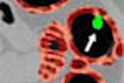
Radiologists like to think that if they find a small spiculated cancer, it means they found it early, meaning they can cure the patient, she said. And often they can. But, showing an image of a tiny nodule invading a vital structure, a pulmonary vein, at presentation, Kazerooni said the size of a nodule isn't always relevant. In a critical location, a small cancer can be unresectable.
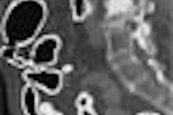
"Not all cancers are nodules in the lung parenchyma," she said. "Many of them are bronchial lesions, particularly squamous cell carcinoma; they begin as a little focal thickening of the bronchial wall and grow into a mass. If we're not even looking at the airway for these things, we're probably missing half of them in screening CT."
When to screen and whom to screen are other issues that are still being resolved. Should risk factors be considered, or family history? What is a significant nodule? Radiologists show significant interreader variability in measuring them, so an accurate 3-D nodule extraction program is needed, Kazerooni said.
Sliding maximum-intensity projection (MIP) is available today. These allow thicker slabs to be seen, which can enable better delineation of blood vessels, and help radiologists distinguish nodules from anatomic structures. On the other hand, rigorous evaluation of the CT data is still tedious and time-consuming, requiring radiologists to scroll in and out of each lobe quadrant in order to see the entire lung.
Computer-aided detection (CAD) could help radiologists find lesions, automatically measure nodules, and automatically calculate their doubling time, but an advanced analysis package isn't available yet.
"The need for computer-aided diagnosis ... is critical," Kazerooni said.
For now, radiologists must find ways to manage the false-positives, and the patients' fears.
"If you're doing thin-section screening CT, and 7 or 8 out of 10 people have a positive finding, then you should probably tell them that up front ...so that when you inform them that they have a nodule they don't have a huge amount of anxiety. "
Individuals' enthusiasm for screening can quickly turn to anxiety when the results are indeterminate, she said, and for now, they have to live with the fear until the nodules are followed up.
Kazerooni is participating in the National Lung Cancer trial, funded by the National Cancer Institute and in cooperation with the PLCO (Prostate, Lung, Colon and Ovarian Cancer Screening Trial). Over 10 years, the trial will screen and follow up on 50,000 smokers and ex-smokers randomly with low-dose chest CT or x-ray in an effort to determine if CT can reduce mortality.
By Eric BarnesAuntMinnie.com staff writer
November 11, 2002
A CD-ROM of the entire SBCT/MR course is available at the organization's Web site.
Related Reading
Pilot study finds that CT lung screening could predict mortality, October 10, 2002
Whole-body CT: Radiology to the people?, October 2, 2002
CT, x-ray to face off in lung cancer screening megatrial, September 19, 2002
Half-dose chest CT produces acceptable image quality, August 5, 2002
Spiral CT promising for lung cancer screening, August 2, 2002
Copyright © 2002 AuntMinnie.com