Even after slashing the radiation dose by half, Canadian investigators were still able to obtain high-quality CT images in patients with renal colic. Dr. James Bell, an abdominal imaging fellow at Vancouver General Hospital in British Columbia, led a study that tested reduced-dose CT in this population. He presented the results at the 2002 American Roentgen Ray Society meeting in Atlanta last month.
Intravenous pyelography (IVP) was the gold standard for diagnosing renal colic for many years, until CT took over, Bell said.
"There are several reasons for this, the most important of which is that CT has, no doubt, an improved sensitivity, specificity, and accuracy over IVP in stone identification. In addition, CT is quicker, requires no IV contrast, and in a significant portion of patients, will identify extraurinary pathology that wouldn’t have been seen on the IVP," he said.
"Despite all these advantages, one of the big disadvantages to CT is that when performed using standard, conventional imaging parameters, it delivers to the patient approximately 2-3 times the dose of an IVP series," Bell added.
Reducing radiation dose in renal colic patients is particularly important because this group tends to be young and of reproductive age.
"(In male patients) the gonads lie near the scan range, and in females, the ovaries are directly radiated," Bell said. "Furthermore, some of the radiological treatment for renal colic involves further exposure to radiation."
The group performed nonenhanced spiral CT in 17 patients with suspected renal colic, using a HiSpeed CT/i (GE Medical Systems,Waukesha, WI). Images were acquired using a slice thickness of 5 mm, pitch of 1.5, and a reduced dose of 120 mAs. Patients were scanned in the prone position, from above the kidney to below the bladder base.
 |
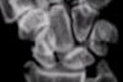
Left ureterovesicle junction stone (UVJ) as seen on the 120-mAs CT scan above and on the 240-mAs scan below.
 |
Six additional images using the conventional dose of 250 mAs also were obtained in each patient at the following levels: left renal hilum, sacral promontory, and mid-pelvis, and at three contiguous levels determined by pathology identified on the complete 120-mAs exam.
Three radiologists specializing in abdominal imaging were blinded to the results and, after reviewing the images, had to reach a consensus. They evaluated six selected 240-mAs scans and six matched 120-mAs scans. A three-point scale was used to assess visualization of the normal anatomic structures; urinary tract pathology, including primary and secondary signs of renal stone disease; other pathology; and image noise.
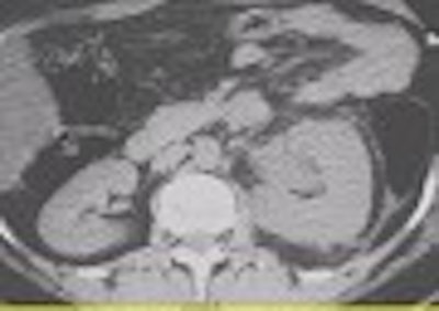
According to the results, visualization of the renal contour, perirenal fat, ureteric contour, and periureteric fat was identical on both the low-dose and conventional-dose scans. In 2 of the 17 patients, renal calculi were seen in both sets of images; in 9 patients, ureteral calculi were diagnosed based on low- and high-dose scans. Finally, a fibroid uterus, right ovarian cyst, and abdominal aortic aneurysm were seen equally well, Bell reported.
 |
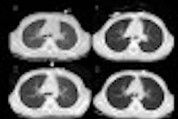
On 120 mAs (above) and 240 mAs (below), an extraurinary diagnosis of right hemorrhagic ovarian cysts and abdominal aortic aneurysm. Images courtesy of Dr. James Bell.
 |
Although the readers reported higher image noise on the 120-mAs images, this evaluation was subjective, and did not alter the fact that the reduced-dose scans were deemed clinically acceptable.
One study limitation was that the group only looked at image quality, not at the diagnostic accuracy of low-dose CT. An ARRS session attendee wondered if lowering dose could mean that smaller stones would be missed. Bell said that the smallest stone his group saw was 1 mm.
However, "using a low-mAs technique still allows good visualization of renal anatomy and pathology," Bell concluded.
Also, the group did not use patient size and weight as criteria for excluding patients from this study. All patients with renal colic were included, and their abdominal size was measured in an AP circumference, he said.
The reduced-dose protocol has been in place at Vancouver General Hospital for over a year and has been successful in large patients, Bell told AuntMinnie.com. Image noise should become less important as readers grow accustomed to looking at lower-dose images, he added.
By Shalmali PalAuntMinnie.com staff writer
May 24, 2002
Related Reading
One-stop evaluation with novel CT urography approach on modified machine, November 27, 2000
Studies show CT vanquishing intravenous urography in flank pain evaluation, May 1, 2000
Copyright © 2002 AuntMinnie.com