CT scanning, by virtue of its capability to image in the axial plane, has proved to be an effective method of documenting injuries. The modality is particularly adept in complex bony anatomy such as the spine, wrist, elbow, and hips. CT arthrography is the standard procedure for the evaluation of ligamentous abnormalities about the wrist when surgical intervention is being considered and plain radiography is nondiagnostic.
Standard CT arthrography protocols for ligament tears call for imaging in two planes, the coronal and transaxial. With the use of high-resolution multislice CT and multiplanar reformatting (MPR), it’s possible to achieve adequate visualization of suspected tears with a single axial acquisition, according to French researchers.
Dr. Sandrine Iochum, from the department of radiology at the Centre Hospitalier Universitaire in Nancy, France, presented the results of her colleagues’ work at the 2001 RSNA meeting in Chicago. The team conducted a side-by-side comparison of coronal and sagittal MPR images versus direct coronal slices in the diagnosis of wrist ligament tears.
The study included 75 patients, ages 17 to 46, presenting with wrist pain. Each patient underwent CT arthrography to depict ligament tears. The exams were performed on a Somatom Volume Zoom multislice CT scanner (Siemens Medical Solutions, Iselin, NJ). The CT arthrography protocol consisted of transaxial and direct coronal slices, 0.5 mm thick, completed with coronal and sagittal MPR, 1 mm thick.
 |
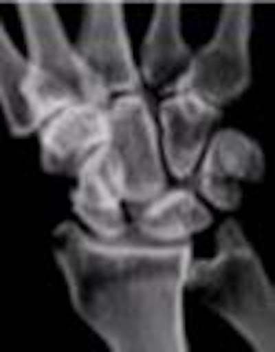
Direct and multiplanar reformatted views of wrist CT arthrography show nearly equivalent information for the depiction of ligament tears. Images courtesy of Dr. Sandrine Iochum, CHU Nancy, France.
 |
The final diagnosis was obtained by consensus of two radiologists working independently. Each of them analyzed two sets of CT slices: axial and direct coronal slices and axial and MPR. The radiologists then assessed the CT arthrographies for scapholunate ligament tears, luno-triquetral ligament tears, and triangular fibrocartilage complex (TFCC) tears. They were also asked to size the TFCC tears (<1 mm, 1-3 mm, and >3 mm).
The researchers found that 35% (26/75) of the patients had a scapholunate tear, 42% (31/75) had a luno-triquetral tear, and 24% (18/75) a TFCC tear. Fourteen of the patients were diagnosed with tears of all three ligaments. The degree of intra and inter-observer reproducibility for the assessment of ligament tears had a kappa value of 0.94 and 0.89, respectively.
There was very close agreement between the two groups of images for the diagnosis of ligament tears. The researchers reported kappa values of 0.96 (direct slices) and 0.98 (MPR) for the scapholunate ligament, 1 and 0.86 for the TFCC, and 0.86 and 0.78 for the luno-triquetral ligament. However, Iochum did note that the degree of agreement for the evaluation of the size of the TFCC tears was not as close, with kappa values of 0.62 and 0.47, respectively.
"We found that CT arthrography with 0.5-mm axial slices from a single axial acquisition provides excellent MPRs for the assessment of wrist ligament tears, and that direct coronal views can be avoided in many situations. The patient undergoing this procedure also benefits from MPR, as reducing the number of views will lower the radiation dose," Iochum said.
By Jonathan S. BatchelorAuntMinnie.com staff writer
March 22, 2002
Related Reading
UK's junior doctors frequently misdiagnose wrist injuries', January 11, 2002
Multislice CT yields simpler method of diagnosing wrist ligament tears, March 7, 2001
Ultrasound and MRI fumble in rheumatoid fingers, March 15, 2000
Copyright © 2002 AuntMinnie.com