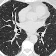
Standardized whole-body CT performed immediately after resuscitation in trauma patients provides better results than a targeted approach using CT, x-ray, or ultrasound, according to a study presented at the 2001 RSNA meeting.
To assess the diagnostic accuracy of a standardized whole-body spiral CT protocol in the initial workup of trauma patients, Dr. Thomas Albrecht and his colleagues from the Universitätsklinikum Benjamin Franklin, Freie Universität in Berlin, Germany, performed a retrospective study on the clinical reports for 46 trauma patients given standardized spiral CT in 2000.
Immediately following resuscitation, the trauma center performed standardized spiral CT exams for the following anatomic regions on each of the 46 patients: neurocranium (unenhanced); skull base (unenhanced; 3-mm collimation, 5-mm/sec table speed, and 3-mm reconstruction interval); chest (150 ml contrast agent, 5-mm collimation, 7.5-mm/sec table speed, and 5-mm reconstruction interval); abdomen (150 ml contrast agent, 5-mm collimation, 7.5-mm/sec table speed, and 5-mm reconstruction interval); and pelvis (150 ml contrast agent, 5-mm collimation, 7.5-mm/sec table speed, and 5-mm reconstruction interval).
The researchers estimated that the center resuscitated patients approximately 45 minutes after their admission to the hospital.
They then compared the CT findings at the initial examination with the final diagnoses at the time of discharge or death. Also, they compared sonography and CT in 22 patients whose records contained reports from an initial abdominal sonogram performed in the intensive care unit.
Final diagnoses included the following injuries: cervical hematoma, hemothorax, pneumothorax, lung contusion, mediastinal hematoma, aortic dissection, retroperitoneal hematoma, renal hematoma and laceration, hepatic hematoma, splenic hematoma and rupture, mesenteric laceration, and isolated intraperitoneal hemorrhage.
Abdominal ultrasound and chest x-ray proved insufficient to exclude common trauma injuries, according to Albrecht.
Spiral CT showed all the injuries listed in the final diagnoses, with the exception of a delayed splenic rupture that occurred several hours after admission in a patient who had developed disseminated intravascular coagulation. Ultrasound missed this splenic rupture as well.
In the 22 patients for whom sonography records were available, the authors found that two hepatic, one splenic, and one renal injury were missed. In addition, sonography resulted in two false-positive findings: one hemopericardium and one renal contusion.
Unlike targeted approaches with CT, x-ray, or ultrasound, whole-body spiral CT provides fast, comprehensive, and accurate diagnosis of cervical, thoracic, and abdominal soft tissue and organ injuries, Albrecht said.
Using whole-body spiral CT, "you’ll find injuries that you do not expect based on clinical evaluation," he said.
By Leslie FarnsworthAuntMinnie contributing writer
January 21, 2002
Related Reading
CT sees growth, and new concerns over radiation dose, November 25, 2001
For the person who has everything, whole-body CT makes inroads, September 11, 2001
Studies and practice shape CT's role in c-spine trauma, August 28, 2001
Abdominal sonography appears useful in triaging patients with penetrating trauma, July 6, 2001
FDA's radiation concerns may lead to dose displays for scanners, May 22, 2001
Copyright © 2002 AuntMinnie.com


