When it comes to working up palpable breast abnormalities with contrast-enhanced mammography (CEM), recombined images better depict the lesion than low-energy ones, research published November 8 in Academic Radiology has found.
The findings shed light on a condition that, although is most commonly benign, can also can be a common presenting symptom of breast cancer, wrote a team led by Tali Amir, MD, of Memorial Sloan Kettering Cancer Center in New York City.
"As such, a thorough work-up [of palpable breast lesions] to exclude carcinoma must always be performed," the group noted.
CEM is a promising technology for this application, the team explained. It consists of a dual-energy digital mammogram performed with iodinated contrast and offers the same anatomic information as a digital mammogram.
Amir and colleagues sought to investigate the role CEM could play in the characterization of palpable breast abnormalities via a study that included 237 women with 262 breast irregularities. The women underwent CEM evaluation, which consisted of the acquisition of low-energy images as well as recombined images (which visualize contrast enhancement) followed by targeted ultrasound. Two readers reviewed the exam findings and made BI-RADS assessments based on low-energy images alone; low-energy plus ultrasound images; recombined images with low-energy plus ultrasound images; and recombined images alone. The investigators used pathology or one-year follow-up imaging as the reference standard.
Of the total number of breast abnormalities, 92% were benign, the team reported. It also found that the recombined images had better specificity compared with the low-energy images in combination with ultrasound:
| Performance of recombined imaging alone with low-energy imaging plus ultrasound for characterizing palpable breast abnormalities | |||
|---|---|---|---|
| Measure | Low-energy imaging plus ultrasound | Recombined imaging alone | p-value |
| Specificity | |||
| Reader 1 | 89% | 94% | p = 0.009 |
| Reader 2 | 88% | 93% | p = 0.03 |
| Positive predictive value | |||
| Reader 1 | 42% | 52% | p = 0.04 |
| Reader 2 | 42% | 53% | p = 0.04 |
| Accuracy | |||
| Reader 1 | 89% | 93% | p = 0.05 |
| Reader 2 | 90% | 93% | p = 0.06 |
"Recombined images had better specificity compared to low energy in combination with ultrasound," the authors concluded. "There was no difference in performance between CEM plus ultrasound and low energy plus ultrasound, likely reflecting the weight ultrasound carries in radiologist decision-making. However, the results indicate that the absence of enhancement on recombined images in the setting of palpable lesions may help avoid benign biopsies."
The complete study can be found here.

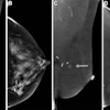

![A normal mammogram confirmed by three-year radiologic follow-up illustrates reader-marked regions of interest (ROIs) during (A) unaided (round 1) and (B) artificial intelligence (AI)–assisted (round 2) reading. Each colored dot represents an ROI for recall by a human reader. Readers could mark more than one ROI per case, represented by multiple dots of the same color. During AI-assisted reading, the AI system displayed three visible prompts: two with suspicion of malignancy scores of 35% (left mediolateral oblique [L MLO] and craniocaudal [L CC]) and one with a suspicion of malignancy score of 10% (right craniocaudal [R CC]), shown as polygonal overlays. Without AI, six of 10 readers (60%) marked a false-positive ROI. With AI assistance, this fell to two of 10 (20%). R MLO = right mediolateral oblique.](https://img.auntminnie.com/mindful/smg/workspaces/default/uploads/2026/07/2026-07-14-radiology-mammogram-ai-auto-bias.H0bYO8QlWs.jpg?auto=format%2Ccompress&fit=crop&h=100&q=70&w=100)


![A normal mammogram confirmed by three-year radiologic follow-up illustrates reader-marked regions of interest (ROIs) during (A) unaided (round 1) and (B) artificial intelligence (AI)–assisted (round 2) reading. Each colored dot represents an ROI for recall by a human reader. Readers could mark more than one ROI per case, represented by multiple dots of the same color. During AI-assisted reading, the AI system displayed three visible prompts: two with suspicion of malignancy scores of 35% (left mediolateral oblique [L MLO] and craniocaudal [L CC]) and one with a suspicion of malignancy score of 10% (right craniocaudal [R CC]), shown as polygonal overlays. Without AI, six of 10 readers (60%) marked a false-positive ROI. With AI assistance, this fell to two of 10 (20%). R MLO = right mediolateral oblique.](https://img.auntminnie.com/mindful/smg/workspaces/default/uploads/2026/07/2026-07-14-radiology-mammogram-ai-auto-bias.H0bYO8QlWs.jpg?auto=format%2Ccompress&dpr=2&fit=crop&h=167&q=70&w=250)


