Wednesday, December 2 | 12:15 p.m.-12:45 p.m. | QS128-ED-WEA3 | Lakeside Learning Center, Station 3
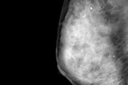
A team from Stony Brook University will detail its success in significantly decreasing the patient no-show rate at a breast imaging center.Anecdotal evidence at the institution's breast imaging center suggested that a large proportion of scheduled exams were not being performed, so an interdisciplinary team of radiologists, technicians, nurses, administrators, and clerical staff was formed with the primary goal of improving workflow and productivity, according to senior author Dr. Nalini Kanth.
An interval review found that the vast majority of unfilled appointments were due to patient no-shows, and interventions were initiated to address this issue, Kanth said. Thanks to systematic preappointment phone calls and documentation by clerical staff, the institution was able to decrease the no-show rate from 21% to 7.1%.
"Getting all key players together in an interdisciplinary team was an important process in identifying the problem and root cause," Kanth told AuntMinnie.com.


![A normal mammogram confirmed by three-year radiologic follow-up illustrates reader-marked regions of interest (ROIs) during (A) unaided (round 1) and (B) artificial intelligence (AI)–assisted (round 2) reading. Each colored dot represents an ROI for recall by a human reader. Readers could mark more than one ROI per case, represented by multiple dots of the same color. During AI-assisted reading, the AI system displayed three visible prompts: two with suspicion of malignancy scores of 35% (left mediolateral oblique [L MLO] and craniocaudal [L CC]) and one with a suspicion of malignancy score of 10% (right craniocaudal [R CC]), shown as polygonal overlays. Without AI, six of 10 readers (60%) marked a false-positive ROI. With AI assistance, this fell to two of 10 (20%). R MLO = right mediolateral oblique.](https://img.auntminnie.com/mindful/smg/workspaces/default/uploads/2026/07/2026-07-14-radiology-mammogram-ai-auto-bias.H0bYO8QlWs.jpg?auto=format%2Ccompress&fit=crop&h=100&q=70&w=100)


![A normal mammogram confirmed by three-year radiologic follow-up illustrates reader-marked regions of interest (ROIs) during (A) unaided (round 1) and (B) artificial intelligence (AI)–assisted (round 2) reading. Each colored dot represents an ROI for recall by a human reader. Readers could mark more than one ROI per case, represented by multiple dots of the same color. During AI-assisted reading, the AI system displayed three visible prompts: two with suspicion of malignancy scores of 35% (left mediolateral oblique [L MLO] and craniocaudal [L CC]) and one with a suspicion of malignancy score of 10% (right craniocaudal [R CC]), shown as polygonal overlays. Without AI, six of 10 readers (60%) marked a false-positive ROI. With AI assistance, this fell to two of 10 (20%). R MLO = right mediolateral oblique.](https://img.auntminnie.com/mindful/smg/workspaces/default/uploads/2026/07/2026-07-14-radiology-mammogram-ai-auto-bias.H0bYO8QlWs.jpg?auto=format%2Ccompress&fit=crop&h=112&q=70&w=112)