Monday, November 30 | 8:50 a.m.-9:00 a.m. | RC215-02 | Arie Crown Theater
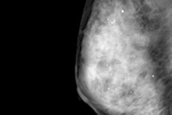
In this session, researchers will describe how screening digital breast tomosynthesis (DBT) offers sustainable and continually improving performance, such as increased cancer detection and fewer interval cancers.Led by Dr. Emily Conant from the University of Pennsylvania Health System, the group sought to examine whether improved screening performance from combining DBT with digital mammography was consistent over consecutive years. To do this, the researchers compared performance metrics from three years of DBT screening, which included more than 33,000 women. They tracked recall rates, cancer detection rates, positive predictive values, biopsy rates, and interval cancer rates.
Conant and colleagues observed the following:
- A 10.4% reduction in the recall rate remained statistically significant over the three-year period.
- Cancer detection rates continued to increase from a baseline rate for digital mammography of 4.6 per 1,000 women to 5.5, 5.8, and 6.1 per 1,000 women for DBT over the three years, although this trend was not statistically significant.
- The biopsy rate remained stable over the three years.
- Interval cancer rates decreased when DBT was used with mammography in year 1 and year 2; the team did not have adequate interval cancer data to calculate the rate for year 3.
The data demonstrate that DBT screening outcomes are sustainable over time and, in fact, continue to improve, the researchers concluded.


![A normal mammogram confirmed by three-year radiologic follow-up illustrates reader-marked regions of interest (ROIs) during (A) unaided (round 1) and (B) artificial intelligence (AI)–assisted (round 2) reading. Each colored dot represents an ROI for recall by a human reader. Readers could mark more than one ROI per case, represented by multiple dots of the same color. During AI-assisted reading, the AI system displayed three visible prompts: two with suspicion of malignancy scores of 35% (left mediolateral oblique [L MLO] and craniocaudal [L CC]) and one with a suspicion of malignancy score of 10% (right craniocaudal [R CC]), shown as polygonal overlays. Without AI, six of 10 readers (60%) marked a false-positive ROI. With AI assistance, this fell to two of 10 (20%). R MLO = right mediolateral oblique.](https://img.auntminnie.com/mindful/smg/workspaces/default/uploads/2026/07/2026-07-14-radiology-mammogram-ai-auto-bias.H0bYO8QlWs.jpg?auto=format%2Ccompress&fit=crop&h=100&q=70&w=100)


![A normal mammogram confirmed by three-year radiologic follow-up illustrates reader-marked regions of interest (ROIs) during (A) unaided (round 1) and (B) artificial intelligence (AI)–assisted (round 2) reading. Each colored dot represents an ROI for recall by a human reader. Readers could mark more than one ROI per case, represented by multiple dots of the same color. During AI-assisted reading, the AI system displayed three visible prompts: two with suspicion of malignancy scores of 35% (left mediolateral oblique [L MLO] and craniocaudal [L CC]) and one with a suspicion of malignancy score of 10% (right craniocaudal [R CC]), shown as polygonal overlays. Without AI, six of 10 readers (60%) marked a false-positive ROI. With AI assistance, this fell to two of 10 (20%). R MLO = right mediolateral oblique.](https://img.auntminnie.com/mindful/smg/workspaces/default/uploads/2026/07/2026-07-14-radiology-mammogram-ai-auto-bias.H0bYO8QlWs.jpg?auto=format%2Ccompress&fit=crop&h=112&q=70&w=112)