Dear AuntMinnie Member,
Our annual Road to RSNA preview of the scientific sessions at RSNA 2015 has gone live!
Road to RSNA gives you a comprehensive look at what will be hot in radiology research in Chicago when RSNA 2015 opens on November 29. The sections are organized by modality, so you can easily find exactly what you want.
We launched Road to RSNA with a preview of the Imaging Informatics sessions; today we're bringing you papers in CT.
More will be coming over the next week and a half, so be sure to check back in our RADCast @ RSNA section at rsna.auntminnie.com.
Regeneration tips for sonographers
In our Ultrasound Community, we're pleased to bring you the latest column in our Making a Difference as a Sonographer series by fitness coach Mark Roozen and echocardiographer Doug Wuebben.
In today's installment, they cover recovery and regeneration tips to achieve an all-around feeling of health and wellness. Adapted from their work in the sports and fitness field, you'll find many of their tips to be just as useful in the often high-stress work that sonographers do.
Just click here to read about it, or go to the Ultrasound Community at ultrasound.auntminnie.com.
DBT finds more cancers
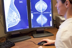
Finally, check out our Women's Imaging Community for a new study that confirms previous research on the effectiveness of digital breast tomosynthesis (DBT) as a screening tool for breast cancer.
Researchers found that DBT not only discovered 54% more cancers than conventional mammography, but it also reduced recall rates. Find out by how much by clicking here, or visit the community at women.auntminnie.com.

![A normal mammogram confirmed by three-year radiologic follow-up illustrates reader-marked regions of interest (ROIs) during (A) unaided (round 1) and (B) artificial intelligence (AI)–assisted (round 2) reading. Each colored dot represents an ROI for recall by a human reader. Readers could mark more than one ROI per case, represented by multiple dots of the same color. During AI-assisted reading, the AI system displayed three visible prompts: two with suspicion of malignancy scores of 35% (left mediolateral oblique [L MLO] and craniocaudal [L CC]) and one with a suspicion of malignancy score of 10% (right craniocaudal [R CC]), shown as polygonal overlays. Without AI, six of 10 readers (60%) marked a false-positive ROI. With AI assistance, this fell to two of 10 (20%). R MLO = right mediolateral oblique.](https://img.auntminnie.com/mindful/smg/workspaces/default/uploads/2026/07/2026-07-14-radiology-mammogram-ai-auto-bias.H0bYO8QlWs.jpg?auto=format%2Ccompress&fit=crop&h=100&q=70&w=100)


![A normal mammogram confirmed by three-year radiologic follow-up illustrates reader-marked regions of interest (ROIs) during (A) unaided (round 1) and (B) artificial intelligence (AI)–assisted (round 2) reading. Each colored dot represents an ROI for recall by a human reader. Readers could mark more than one ROI per case, represented by multiple dots of the same color. During AI-assisted reading, the AI system displayed three visible prompts: two with suspicion of malignancy scores of 35% (left mediolateral oblique [L MLO] and craniocaudal [L CC]) and one with a suspicion of malignancy score of 10% (right craniocaudal [R CC]), shown as polygonal overlays. Without AI, six of 10 readers (60%) marked a false-positive ROI. With AI assistance, this fell to two of 10 (20%). R MLO = right mediolateral oblique.](https://img.auntminnie.com/mindful/smg/workspaces/default/uploads/2026/07/2026-07-14-radiology-mammogram-ai-auto-bias.H0bYO8QlWs.jpg?auto=format%2Ccompress&fit=crop&h=112&q=70&w=112)