Physicians at Memorial Sloan-Kettering Cancer Center in New York City have developed a predictability model for determining the risk of ipsilateral breast tumor recurrence after breast conservation surgery for women diagnosed with early-stage, low-risk ductal carcinoma in situ (DCIS).
Using this tool to calculate the individual risk of a patient five and 10 years after surgery may assist both the patient and her physician in evaluating treatment options, according to an article published online in the Journal of Clinical Oncology (July 12, 2010).
In the U.S. alone, an estimated 54,010 cases of breast carcinoma in situ are expected to be diagnosed in 2010, according to statistics from the American Cancer Society (ACS). Although clinical trials show that adjuvant radiation therapy and antiestrogen therapy decrease the rate of ipsilateral breast tumor recurrence by approximately 50% and 30%, respectively, some clinicians and some patients question whether the toxicities and potential morbidity of adjuvant treatment are an appropriate trade-off risk in view of the overall low risk of cancer recurrence.
 |
| Click here to enlarge this image. |
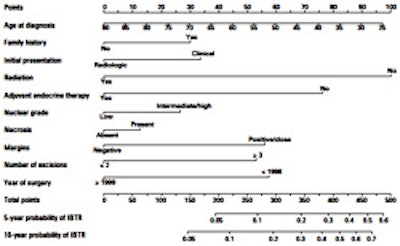
| Nomogram for predicting five- and 10-year probability of ipsilateral breast tumor recurrences after breast conserving surgery for DCIS. Published with permission of the Journal of Clinical Oncology. |
This prediction model, a nomogram, combines 10 factors shown to affect risk. Users calculate the total number of points for 10 clinical and pathological variables, and draw a vertical line from the total point line to the five- and 10-year local recurrent lines.
To develop the model, Kimberly Van Zee, MD, a surgical oncologist at Memorial Sloan-Kettering's Evelyn H. Lauder Breast Cancer Center, and colleagues evaluated the clinicopathologic characteristics of 1,868 consecutive patients treated with breast conservation surgery for DCIS between 1991 and 2006.
Data from this patient population, excluding 187 women who either did not have a pure DCIS diagnosis or a complete record, were analyzed to develop the nomogram.
Out of the total patient population, 202 patients, or 10.8%, had a cancer recurrence; 40% had a recurrence with invasive breast cancer, and 60% had recurrence with DCIS.
The risk estimation tool should be used in conjunction with an assessment of a patient's overall health and competing risk of death from other causes, the authors cautioned.
By Cynthia E. Keen
AuntMinnie.com staff writer
July 29, 2010
Related Reading
Elderly early-stage breast cancer patients don't need RT, May 21, 2010
More frequent follow-up helps breast conservation patients, May 5, 2010
DCIS recurrence rates higher in younger women, October 7, 2009
Combination surgery and radiotherapy benefits all women with DCIS, February 9, 2009
Copyright © 2010 AuntMinnie.com