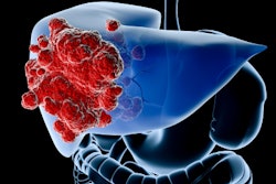
Microbubble ultrasound contrast agents improve liver tumor response to radiotherapy and thus increase patient survival, researchers have reported.
In two studies, a team led by John Eisenbrey, PhD, of Thomas Jefferson University in Philadelphia found that patients with aggressive intrahepatic cholangiocarcinoma (ICC) and liver metastasis showed a greater than 50% improvement in tumor response rates and that patients with hepatocellular carcinoma (HCC) experienced "significant improvement on both tumor response and patient survival when radiotherapy embolization was enhanced using a microbubble ultrasound contrast agent and ultrasound." The group's results were presented on September 10 at the International Contrast Ultrasound Society (ICUS) meeting in Chicago.
Standard radioembolization therapy for liver cancer uses glass beads filled with the radioactive isotope yttrium-90 (Y-90), which are distributed to the tumor via a catheter. In particular, ICC tumors tend to be aggressive and have a poor prognosis, Eisenbrey said, noting, however, that although prior studies have shown favorable results for CEUS-enhanced radiotherapy in patients with breast and head and neck cancer, "these [studies] are the first trials showing safety and feasibility for liver cancer."
"Standard CEUS utilizes microbubble ultrasound contrast agents (UCAs) to enhance diagnostic ultrasound images, but UCA uses are now expanding to therapy, … [and] UCAs do not contain iodine or gadolinium and are not nephrotoxic," he said.







![Examples of ultrasound findings and techniques. (A) Images in a 39-year-old male patient with a mass in the left thigh. The mass is heterogeneous on the B-mode US image (compared with the patient in D) and showed increased microvascularity (superb microvascular imaging [SMI]) and shear-wave elastography (SWE) values. Undifferentiated pleomorphic sarcoma was diagnosed at biopsy (with pleomorphic rhabdomyosarcoma in surgical specimen). (B) Images in an 18-year-old male patient with a mass in the left leg. The mass is hypoechoic on the B-mode image, with no other findings suggestive of malignancy. The lesion is in contact with the cortex of the tibia, which is slightly irregular. CT revealed a doubtful anteromedial tibial erosion. The microvascular study demonstrated high vascularization, suggestive of malignancy. Periosteal Ewing sarcoma was diagnosed with both histologic and immunohistochemical confirmation. (C) Images in a 69-year-old female patient with a lump growing on the outside of the left leg. Multiple SWE examinations were performed (please note the high values obtained in the measurements, whereas the color map highlights the stiffness relative to adjacent tissues). SMI showed areas of increased vascularization to target for sampling. Undifferentiated spindle cell sarcoma was diagnosed at biopsy, with residual leiomyosarcoma in the surgical specimen after neoadjuvant therapy. (D) Images in a 56-year-old female patient with a mass in the right thigh. The mass is heterogeneous at both B-mode ultrasound (similar to patient A) and MRI (coronal T2-weighted spectral attenuated inversion recovery [SPAIR]; T1-weighted pre-contrast and postcontrast imaging), which even shows uptake after the administration of paramagnetic contrast material, which is traditionally suggestive of malignancy. Low values at SMI and elastography are suggestive of benignity. Spindle cell lipoma was diagnosed at biopsy, with atypical spindle cell lipomatous tumor in the surgical specimen.](https://img.auntminnie.com/mindful/smg/workspaces/default/uploads/2026/08/images-radiol250278fig2.APCFLSvX6p.jpg?auto=format%2Ccompress&fit=crop&h=112&q=70&w=112)


