AI improves detection of major congenital heart defects (CHDs) on prenatal ultrasound, suggest findings presented January 30 at the Annual Pregnancy Meeting of the Society for Maternal-Fetal Medicine (SMFM) in Denver, CO.
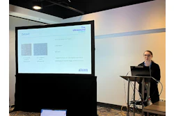
In her presentation, Jennifer Lam-Rachlin, MD, from Mount Sinai West in New York presented her team’s findings, showing that adding AI to prenatal ultrasound exams has several benefits, including improved performance and confidence among ob/gyn and maternal-fetal medicine (MFM) practitioners, as well as decreased evaluation times.
“AI can be a tremendous ultrasound screening aid for major congenital heart defects,” Lam-Rachlin said.
Congenital heart defects occur in between 1% and 2% of all pregnancies. Of these cases, around one in four consist of severe or critical heart conditions.
Prenatal detection rates on ultrasound vary around the world between 30% and 50%, owing to variability among medical centers. This includes many screening anatomy ultrasounds not being performed by expert sonographers, Lam-Rachlin said.
Previous research suggests that AI can aid novice ultrasound users. A 2025 study led by Yale University researchers found that novice users of lung ultrasound (LUS) can successfully acquire diagnostic-quality images with AI assistance.
Using an AI screening tool (BrightHeart), Lam-Rachlin and colleagues investigated whether AI help can improve CHD detection on fetal ultrasound exams among both ob/gyns and MFM specialists.
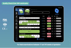
 Example of an ultrasound exam being evaluated through an AI system. Researchers found that AI assistance boosted CHD performance among ob/gyns and MFMs reading ultrasound exams.Mount Sinai West
The AI system analyzes grayscale 2D ultrasound exams and detects eight morphological findings associated with severe CHDs. The team used a dataset of 200 ultrasound exams from 11 centers in two countries, with 100 exams having at least one suspicious finding. The researchers also noted that these exams were not used to train the AI system. A panel of experienced fetal cardiologists provided ground truth analysis.
For the study, 14 ob/gyns and MFMs with varying experience (one year to more than 30 years) reviewed each ultrasound exam both aided and unaided by the AI system in randomized order. From there, they annotated the exams for the presence or absence of CHD findings.
| Performance of physicians in reading prenatal ultrasound exams with and without AI assistance | ||
|---|---|---|
| Measure | Unassisted reading | AI-assisted reading |
| Receiver operator characteristics area under the curve (ROC AUC) | 0.83 | 0.97 |
| Sensitivity | 78% | 94% |
| Specificity | 76% | 97% |
| Reading time | 274 seconds | 226 seconds |
| *All results achieved statistical significance. | ||
The team also highlighted that the AI system improved performance regardless of patient characteristics or exam quality. And the physicians’ confidence significantly improved with AI assistance from 3.9 to 4.6 (p < 0.001).
And while AI assistance trended toward improvement for all levels of experience, physicians with less than three years of experience benefitted the most, with the ROC AUC improving from 0.8 to 0.98 (p = 0.05).
Lam-Rachlin said that with these results in mind, AI help could improve neonatal mortality and morbidity rates, though more research will be needed.
She added that prospective studies are needed to evaluate AI-assistance software in real-world clinical settings and its performance in underresourced areas.