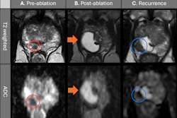
MRI-guided transurethral ultrasound ablation (TULSA) is effective over surgery in men with prostate cancer, according to a study presented April 13 at the Society of Interventional Radiology (SIR) annual meeting in Toronto.
The finding is from a clinical trial comparing TULSA to robotic prostatectomy in 212 men at 23 medical centers between 2022 and 2025, noted principal investigator David Woodrum, MD, PhD, of the Mayo Clinic in Rochester, MN.
"These early results suggest that TULSA may allow patients to recover more quickly and maintain a better quality of life following treatment, while still effectively treating the cancer," Woodrum said, in a news release from SIR.
TULSA uses real-time MRI to guide the delivery of high-energy ultrasound through the urethra and into the prostate to heat and destroy cancer tissue without affecting surrounding organs, with the goal of preserving urinary and sexual function. Robotic prostatectomy, while effective for cancer control, can leave men with long-term effects including erectile dysfunction and urinary incontinence, Woodrum explained.
In the CAPTAIN trial, participants were randomly assigned to receive either TULSA or robotic prostatectomy. For TULSA versus surgery participants, the median age was 63 years old versus 65, median PSA was 6.5 ng/mL versus 7.2 ng/mL, and prostate volume was 41 cc versus 35 cc.
According to the results, men treated with TULSA had less blood loss during the procedure (zero mL versus 100 mL to 200 mL), had shorter stays (0.29 days versus 1.24 days), and reported less pain and faster return to normal activities one month after treatment.
"For many patients, how quickly they can get back to work, family life and everyday routines really matters," Woodrum said.
The researchers plan to follow participants over 10 years to compare longer-term outcomes, including urinary control, sexual function, and whether additional cancer treatment is needed.







![Examples of ultrasound findings and techniques. (A) Images in a 39-year-old male patient with a mass in the left thigh. The mass is heterogeneous on the B-mode US image (compared with the patient in D) and showed increased microvascularity (superb microvascular imaging [SMI]) and shear-wave elastography (SWE) values. Undifferentiated pleomorphic sarcoma was diagnosed at biopsy (with pleomorphic rhabdomyosarcoma in surgical specimen). (B) Images in an 18-year-old male patient with a mass in the left leg. The mass is hypoechoic on the B-mode image, with no other findings suggestive of malignancy. The lesion is in contact with the cortex of the tibia, which is slightly irregular. CT revealed a doubtful anteromedial tibial erosion. The microvascular study demonstrated high vascularization, suggestive of malignancy. Periosteal Ewing sarcoma was diagnosed with both histologic and immunohistochemical confirmation. (C) Images in a 69-year-old female patient with a lump growing on the outside of the left leg. Multiple SWE examinations were performed (please note the high values obtained in the measurements, whereas the color map highlights the stiffness relative to adjacent tissues). SMI showed areas of increased vascularization to target for sampling. Undifferentiated spindle cell sarcoma was diagnosed at biopsy, with residual leiomyosarcoma in the surgical specimen after neoadjuvant therapy. (D) Images in a 56-year-old female patient with a mass in the right thigh. The mass is heterogeneous at both B-mode ultrasound (similar to patient A) and MRI (coronal T2-weighted spectral attenuated inversion recovery [SPAIR]; T1-weighted pre-contrast and postcontrast imaging), which even shows uptake after the administration of paramagnetic contrast material, which is traditionally suggestive of malignancy. Low values at SMI and elastography are suggestive of benignity. Spindle cell lipoma was diagnosed at biopsy, with atypical spindle cell lipomatous tumor in the surgical specimen.](https://img.auntminnie.com/mindful/smg/workspaces/default/uploads/2026/08/images-radiol250278fig2.APCFLSvX6p.jpg?auto=format%2Ccompress&fit=crop&h=112&q=70&w=112)


