Contrast-enhanced ultrasound (CEUS) may be preferred for finding enhancement in indeterminate kidney lesions, suggest findings published February 12 in WFUMB Ultrasound Open.
CEUS showed near-perfect interobserver agreement and superior sensitivity compared to color Doppler ultrasound in this area, wrote a team led by Madeleine Sertic, MD, from Massachusetts General Hospital in Boston.
“All biopsy-proven renal neoplasms enhanced on CEUS, including those without detectable Doppler flow,” Sertic and colleagues wrote.
Conventional B-mode ultrasound and color Doppler imaging struggle to differentiate between benign and malignant kidney lesions, especially when the lesions are small, cystic, or show low-flow microvascularity. This leads to indeterminate lesions being further evaluated via cross-sectional imaging, biopsy, or unnecessary nephrectomy.
CEUS could help in this area by assessing real-time tissue perfusion with intravenously administered microbubble contrast agents, the researchers noted. And prior research focusing on CEUS in renal imaging suggests moderate to good interobserver reproducibility.
Sertic and co-authors compared inter-reader agreement for color Doppler vascularity and CEUS enhancement in indeterminate kidney lesions. They also compared the sensitivity of each technique in characterizing kidney masses.
The study included 38 patients who underwent CEUS evaluation at two tertiary care centers between 2019 and 2025. Two board-certified, subspecialty-trained radiologists independently assessed color Doppler vascularity and enhancement using sulfur hexafluoride microbubble contrast. They were blinded to final diagnoses.
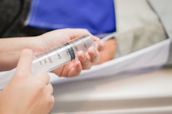
 Images show an enhancing mass with no detectable flow on color Doppler. (A) Sagittal B-mode ultrasound in a 71-year-old male with an incidental right renal mass reveals a hyperechoic solid renal mass (solid arrow), with no flow on color Doppler and an adjacent simple cyst (dashed arrow). (B) Split-screen image following contrast injection during CEUS reveals heterogeneous enhancement of the solid mass (solid arrow). The simple cyst is nonenhancing (dashed arrow). This solid mass is a presumed neoplasm and is followed with annual surveillance.Creative Commons license (CC BY 4.0)
Final analysis included 45 renal lesions, 12 being solid and 33 being cystic. The team reported an average maximum lesion diameter of 32 mm, with a range of 8 mm to 99 mm.
It also reported strong inter-reader agreement for the assessment of vascularity (Kappa = 0.738) and presence of enhancement (Kappa = 0.814). Lesions were more likely to enhance (n = 34) than show vascularity (n = 13; p < 0.001. And seven enhancing lesions (21%) showed vascularity.
Also, the researchers found nine biopsied lesions that were neoplasms, including eight being renal cell carcinoma and one being oncocytoma. All nine neoplasms showed enhancement on CEUS, indicating 100% sensitivity. However, only five showed vascularity on color Doppler imaging (56% sensitivity).
The results support integrating CEUS into routine diagnostic algorithms for characterizing indeterminate renal lesions, the study authors highlighted.
“CEUS may be an effective strategy to mitigate variability that has been historically associated with the ultrasound modality,” they wrote.
The authors also outlined the following advantages CEUS has over CT and MRI: lack of contrast excretion into the collecting system, lack of ionizing radiation, absence of nephrotoxicity, real-time dynamic assessment, and the ability to perform multiple injections during a single exam.
“These benefits are particularly relevant for patients with renal impairment, contrast allergies, or those requiring serial imaging surveillance,” they added.
Read the full study here.