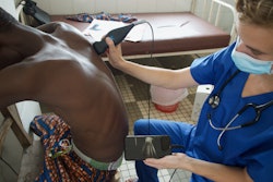
Bedside ultrasound can lessen anxiety and pain for emergency patients being diagnosed with acute cholecystitis, a study published January 8 in the Journal of Emergency Nursing found.
Compared with emergency patients receiving standard care, patients examined via point-of-care ultrasound (POCUS) experienced decreases in anxiety, according to research led by Shaofeng Wang, MD, and Nansheng Cheng, MD, from Sichuan University in China.
“Implementation of point-of-care ultrasound may improve patient experience and potentially enhance clinical outcomes in the management of acute cholecystitis,” Wang and Cheng wrote.
Acute cholecystitis typically occurs when a gallstone blocks the cystic duct, leading to the gallbladder becoming inflamed. Patients usually undergo clinical assessment followed by laboratory testing and radiological confirmation. Ultrasound is used as a first-line imaging method for the latter step. However, this approach requires patients to be transported from the emergency department to the radiology suite, which may lead to delays in diagnosis and treatment.
Prior research has explored the utility of POCUS performed at the bedside, focusing on diagnostic accuracy and time-to-diagnosis in various acute conditions.
Wang and Cheng evaluated the use of bedside POCUS, comparing results with those of standard ultrasound. The study centered around anxiety and pain levels in patients with suspected acute cholecystitis in the emergency department.
The observational study included 100 patients, with 50 undergoing bedside POCUS imaging and the other 50 receiving standard care. The researchers measured patient anxiety with the state-trait anxiety inventory state anxiety subscale (range, 20 to 80) and measured pain by using a visual analog scale (range, 0 to 10). Higher scores for both measures mean elevated levels of anxiety and pain.
The POCUS group showed significantly lower anxiety levels and lower pain scores compared with the standard care group.
Comparison between POCUS, standard care groups | |||
Scores | Standard care group | POCUS group | p-value |
Anxiety score | 48.7 | 42.2 | 0.009 |
Pain score | 6.5 | 5 | 0.0003 |
And while the proportion of patients with high anxiety remained unchanged in the standard care group, the POCUS group experienced a large decrease from 60% (n = 30) to 24% (n = 12).
The study authors suggested that bedside POCUS could improve overall experience and satisfaction for patients with biliary disease when used as a standard component of care. They added that clinical outcomes could be improved, since high anxiety is tied to increased pain perception, longer recovery times, and poorer response to treatment.
“As emergency departments continue to seek effective nonpharmacologic approaches to pain and anxiety management, bedside ultrasound represents a promising intervention that merits consideration for incorporation into standard care protocols for patients with acute cholecystitis,” Wang and Cheng wrote.
Read the full study here.


![Examples of ultrasound findings and techniques. (A) Images in a 39-year-old male patient with a mass in the left thigh. The mass is heterogeneous on the B-mode US image (compared with the patient in D) and showed increased microvascularity (superb microvascular imaging [SMI]) and shear-wave elastography (SWE) values. Undifferentiated pleomorphic sarcoma was diagnosed at biopsy (with pleomorphic rhabdomyosarcoma in surgical specimen). (B) Images in an 18-year-old male patient with a mass in the left leg. The mass is hypoechoic on the B-mode image, with no other findings suggestive of malignancy. The lesion is in contact with the cortex of the tibia, which is slightly irregular. CT revealed a doubtful anteromedial tibial erosion. The microvascular study demonstrated high vascularization, suggestive of malignancy. Periosteal Ewing sarcoma was diagnosed with both histologic and immunohistochemical confirmation. (C) Images in a 69-year-old female patient with a lump growing on the outside of the left leg. Multiple SWE examinations were performed (please note the high values obtained in the measurements, whereas the color map highlights the stiffness relative to adjacent tissues). SMI showed areas of increased vascularization to target for sampling. Undifferentiated spindle cell sarcoma was diagnosed at biopsy, with residual leiomyosarcoma in the surgical specimen after neoadjuvant therapy. (D) Images in a 56-year-old female patient with a mass in the right thigh. The mass is heterogeneous at both B-mode ultrasound (similar to patient A) and MRI (coronal T2-weighted spectral attenuated inversion recovery [SPAIR]; T1-weighted pre-contrast and postcontrast imaging), which even shows uptake after the administration of paramagnetic contrast material, which is traditionally suggestive of malignancy. Low values at SMI and elastography are suggestive of benignity. Spindle cell lipoma was diagnosed at biopsy, with atypical spindle cell lipomatous tumor in the surgical specimen.](https://img.auntminnie.com/mindful/smg/workspaces/default/uploads/2026/08/images-radiol250278fig2.APCFLSvX6p.jpg?auto=format%2Ccompress&fit=crop&h=100&q=70&w=100)


![Examples of ultrasound findings and techniques. (A) Images in a 39-year-old male patient with a mass in the left thigh. The mass is heterogeneous on the B-mode US image (compared with the patient in D) and showed increased microvascularity (superb microvascular imaging [SMI]) and shear-wave elastography (SWE) values. Undifferentiated pleomorphic sarcoma was diagnosed at biopsy (with pleomorphic rhabdomyosarcoma in surgical specimen). (B) Images in an 18-year-old male patient with a mass in the left leg. The mass is hypoechoic on the B-mode image, with no other findings suggestive of malignancy. The lesion is in contact with the cortex of the tibia, which is slightly irregular. CT revealed a doubtful anteromedial tibial erosion. The microvascular study demonstrated high vascularization, suggestive of malignancy. Periosteal Ewing sarcoma was diagnosed with both histologic and immunohistochemical confirmation. (C) Images in a 69-year-old female patient with a lump growing on the outside of the left leg. Multiple SWE examinations were performed (please note the high values obtained in the measurements, whereas the color map highlights the stiffness relative to adjacent tissues). SMI showed areas of increased vascularization to target for sampling. Undifferentiated spindle cell sarcoma was diagnosed at biopsy, with residual leiomyosarcoma in the surgical specimen after neoadjuvant therapy. (D) Images in a 56-year-old female patient with a mass in the right thigh. The mass is heterogeneous at both B-mode ultrasound (similar to patient A) and MRI (coronal T2-weighted spectral attenuated inversion recovery [SPAIR]; T1-weighted pre-contrast and postcontrast imaging), which even shows uptake after the administration of paramagnetic contrast material, which is traditionally suggestive of malignancy. Low values at SMI and elastography are suggestive of benignity. Spindle cell lipoma was diagnosed at biopsy, with atypical spindle cell lipomatous tumor in the surgical specimen.](https://img.auntminnie.com/mindful/smg/workspaces/default/uploads/2026/08/images-radiol250278fig2.APCFLSvX6p.jpg?auto=format%2Ccompress&fit=crop&h=112&q=70&w=112)