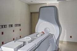
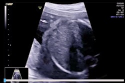
Cranial ultrasound could have utility in imaging infants with hypoxic-ischemic encephalopathy (HIE), suggest findings published November 6 in Pediatric Neurology.
Researchers led by Aine Fox, PhD, of Rotunda Hospital in Dublin, Ireland, found correlations of brain injury findings between cranial ultrasound and brain MRI, which suggest ultrasound may be a suitable alternative in places where MRI access is difficult.
“Early postnatal cranial ultrasound can provide information on potential findings on brain MRI and may help inform outcome of newborns in low-middle income countries and situations where MRI is not clinically possible,” Fox and co-authors wrote.
Access to brain MRI is increasing in high-income countries, putting cranial ultrasound on the back burner for neuroimaging. Current recommendations do not support the use of cranial ultrasound for assessing brain injury in HIE. A previous report also suggested that HIE incidence is significantly lower in high-income countries.
Fox and colleagues study correlations of brain injury findings on early postnatal cranial ultrasound with findings on neonatal brain MRI in a routine clinical setting.
The study included 94 infants with moderate to severe HIE. Sonographers performed cranial ultrasound in the first five days after birth while MRI technologists performed brain MRI exams at a median of 6.7 days.
The team observed that white matter injury findings on ultrasound within 24 hours and gray matter injury on ultrasound within 48 hours correlated with similar nature and severity of brain injury findings on MRI. On subgroup analysis, the researchers found stronger correlations of brain injury between findings on ultrasound performed within 24 hours and after 48 hours and contemporaneous brain MRI performed on days three through five.
| Correlations between ultrasound, MRI findings in infants with HIE | ||||
|---|---|---|---|---|
| Cranial ultrasound performed within 24 hours | Cranial ultrasound performed after 48 hours | |||
| Type of injury | Correlation (Rho) | p-value | Correlation (Rho) | p-value |
| All brain MRI | ||||
| White matter | 0.334 | 0.015 | 0.25 | 0.053 |
| Gray matter | 0.117 | 0.404 | 0.404 | 0.001 |
| Contemporaneous brain MRI | ||||
| White matter | 0.783 | 0.003 | 0.59 | 0.034 |
| Gray matter | 0.272 | 0.392 | 0.609 | 0.027 |
Also, cranial ultrasound achieved a specificity of 91% for finding gray matter injuries on brain MRI. The researchers additionally found that no gray matter injuries on brain MRI were related to a good likelihood of having no gray matter injuries observed on cranial ultrasound. This included negative predictive values (NPVs) of 71% for ultrasounds performed within 24 hours and 78% for ultrasounds performed after 48 hours.
Finally, for infants who experienced a sentinel event, ultrasound exams performed after 48 hours achieved a specificity of 81% for gray matter injuries. And where gray matter injuries were not present on cranial ultrasound, they were not likely to be found on MRI (NPV = 81%).
These findings may help inform neurodevelopmental outcomes for affected newborns in low- to middle-income countries, the study authors highlighted. They further wrote that cranial ultrasound is easily accessible and cost-effective.
“A larger prospective study evaluating the correlation for findings of brain injury between neuroimaging modalities at different time points after birth could better inform the utility of cranial ultrasound in newborns with HIE,” the authors wrote.
The full study can be found here.