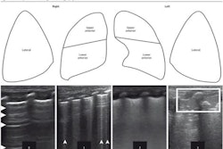
Lung ultrasound scoring can be used to monitor respiratory conditions in preterm infants, according to research published June 7 in JAMA Network Open.
A team led by Luca Bonadies, MD, PhD, from Padova University Hospital in Italy found that infants with later respiratory problems show signs of worse lung aeration as measured by lung ultrasound in the first 14 days of life.
“Good timing to estimate respiratory outcomes seems to be the second week of life, although the ideal day of life remains unknown,” the Bonadies team wrote.
There is growing evidence that lung ultrasound scoring can be used to measure the risk of developing bronchopulmonary dysplasia. Poor respiratory outcomes and frequent respiratory exacerbations are common complications of preterm birth. The researchers however noted a lack of data tying the use of lung ultrasound with later respiratory outcomes.
Bonadies and colleagues studied the association between lung ultrasound within the first three, seven, and 14 days of life, and respiratory outcomes at 2 years corrected age. Outcomes included respiratory exacerbations, physician-diagnosed wheezing episodes, and respiratory medication use.
The study enrolled 51 children with an average gestational age of 27.3 weeks between 2019 and 2022. Of the total, 20 were female and 31 were male, and the infants had an average birth weight of 947 grams. Of the total cohort, 16 (31%) developed moderate to severe bronchopulmonary dysplasia, while eight (16%) were determined to have mild bronchopulmonary dysplasia.
After initial discharge, 32 infants (63%) received at least one respiratory drug, which included bronchodilators, corticosteroids, and/or antibiotics. Sixteen infants (31%) were hospitalized for respiratory problems, and three needed pediatric intensive care unit (ICU) admission. The team reported that the average respiratory morbidity score at two years was 0.51. It also found that infants needing respiratory drugs had a higher median lung ultrasound score compared to those who did not receive drugs or hospitalization.
| Comparison of median lung ultrasound scores between infants needing, not needing drugs or hospitalization | |||
|---|---|---|---|
| Day of life |
Infants not needing drugs | Infants needing drugs |
p-value |
| 3 |
1 |
6 |
0.02 |
| 7 |
0 |
5 |
< 0.001 |
| 14 |
1 |
6 |
< 0.001 |
| Infants not needing hospitalization | Infants needing hospitalization | ||
| 3 |
3 |
4 |
< 0.001 |
| 7 |
1 |
10 |
< 0.001 |
| 14 |
2 |
8 |
< 0.001 |
Infants needing ICU always had lung ultrasound scores over eight, the researchers reported.
Also, lung ultrasound scores correlated with the number of respiratory exacerbations needing drugs at days of life 3, 7, and 14. For example, at day of life 7, the correlation for needing drugs was 0.762, while correlation with hospitalization was 0.663. The team noted the same trends with respiratory morbidity score (ρ [rho] = 0.714), and after correction for gestational age for days of life 7 and 14 (needing drugs, adjusted β = 0.352 and 0.177, respectively).
Finally, the authors found that lung ultrasound scoring was associated with respiratory outcomes. At day of life 7, this resulted in area under the curve (AUC) values of 0.838 for needing drugs, and 0.868 for needing hospitalizations.
The study authors highlighted that while the sample size was small, the results suggest the presence of an active evolving disease in the lungs toward a higher lung ultrasound score in infants later affected.
“Lung ultrasonography performed at day of life three may not be associated with long-term outcomes because lung ultrasound scoring could be altered by other pathological processes, such as respiratory distress syndrome and patent ductus arteriosus,” they added.
The full study can be found here.

![Examples of ultrasound findings and techniques. (A) Images in a 39-year-old male patient with a mass in the left thigh. The mass is heterogeneous on the B-mode US image (compared with the patient in D) and showed increased microvascularity (superb microvascular imaging [SMI]) and shear-wave elastography (SWE) values. Undifferentiated pleomorphic sarcoma was diagnosed at biopsy (with pleomorphic rhabdomyosarcoma in surgical specimen). (B) Images in an 18-year-old male patient with a mass in the left leg. The mass is hypoechoic on the B-mode image, with no other findings suggestive of malignancy. The lesion is in contact with the cortex of the tibia, which is slightly irregular. CT revealed a doubtful anteromedial tibial erosion. The microvascular study demonstrated high vascularization, suggestive of malignancy. Periosteal Ewing sarcoma was diagnosed with both histologic and immunohistochemical confirmation. (C) Images in a 69-year-old female patient with a lump growing on the outside of the left leg. Multiple SWE examinations were performed (please note the high values obtained in the measurements, whereas the color map highlights the stiffness relative to adjacent tissues). SMI showed areas of increased vascularization to target for sampling. Undifferentiated spindle cell sarcoma was diagnosed at biopsy, with residual leiomyosarcoma in the surgical specimen after neoadjuvant therapy. (D) Images in a 56-year-old female patient with a mass in the right thigh. The mass is heterogeneous at both B-mode ultrasound (similar to patient A) and MRI (coronal T2-weighted spectral attenuated inversion recovery [SPAIR]; T1-weighted pre-contrast and postcontrast imaging), which even shows uptake after the administration of paramagnetic contrast material, which is traditionally suggestive of malignancy. Low values at SMI and elastography are suggestive of benignity. Spindle cell lipoma was diagnosed at biopsy, with atypical spindle cell lipomatous tumor in the surgical specimen.](https://img.auntminnie.com/mindful/smg/workspaces/default/uploads/2026/08/images-radiol250278fig2.APCFLSvX6p.jpg?auto=format%2Ccompress&fit=crop&h=100&q=70&w=100)


![Examples of ultrasound findings and techniques. (A) Images in a 39-year-old male patient with a mass in the left thigh. The mass is heterogeneous on the B-mode US image (compared with the patient in D) and showed increased microvascularity (superb microvascular imaging [SMI]) and shear-wave elastography (SWE) values. Undifferentiated pleomorphic sarcoma was diagnosed at biopsy (with pleomorphic rhabdomyosarcoma in surgical specimen). (B) Images in an 18-year-old male patient with a mass in the left leg. The mass is hypoechoic on the B-mode image, with no other findings suggestive of malignancy. The lesion is in contact with the cortex of the tibia, which is slightly irregular. CT revealed a doubtful anteromedial tibial erosion. The microvascular study demonstrated high vascularization, suggestive of malignancy. Periosteal Ewing sarcoma was diagnosed with both histologic and immunohistochemical confirmation. (C) Images in a 69-year-old female patient with a lump growing on the outside of the left leg. Multiple SWE examinations were performed (please note the high values obtained in the measurements, whereas the color map highlights the stiffness relative to adjacent tissues). SMI showed areas of increased vascularization to target for sampling. Undifferentiated spindle cell sarcoma was diagnosed at biopsy, with residual leiomyosarcoma in the surgical specimen after neoadjuvant therapy. (D) Images in a 56-year-old female patient with a mass in the right thigh. The mass is heterogeneous at both B-mode ultrasound (similar to patient A) and MRI (coronal T2-weighted spectral attenuated inversion recovery [SPAIR]; T1-weighted pre-contrast and postcontrast imaging), which even shows uptake after the administration of paramagnetic contrast material, which is traditionally suggestive of malignancy. Low values at SMI and elastography are suggestive of benignity. Spindle cell lipoma was diagnosed at biopsy, with atypical spindle cell lipomatous tumor in the surgical specimen.](https://img.auntminnie.com/mindful/smg/workspaces/default/uploads/2026/08/images-radiol250278fig2.APCFLSvX6p.jpg?auto=format%2Ccompress&fit=crop&h=112&q=70&w=112)