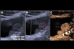
Intussusception reduction can be performed within eight hours of ultrasound diagnosis, not necessarily within two hours, according to research published September 4 in the American Journal of Roentgenology.
A team led by Livja Mertiri, MD, from Texas Children's Hospital in Houston found comparable success for pediatric intussusception reduction in children who were treated emergently (within 2 hours) and those who were treated urgently (within 8 hours).
“The findings have implications for determining the standard of care, including criteria for on-call activation of radiologic resources, in pediatric intussusception management,” Mertiri and colleagues wrote.
Radiologists follow the rule of thumb that pediatric ileocolic intussusceptions should be treated emergently, with delayed reduction attempts potentially contributing to worse health outcomes. But the researchers pointed out that the relevant literature in this area is conflicting.
Mertiri et al identified factors tied to successful image-guided ileocolic intussusception reduction in children. They paid close attention to the time interval since diagnosis.
Final analysis included data collected between 2009 and 2023 from 1,065 patients, all of whom were younger than six years. Of the total, 649 were male and 416 were female with an average age of 18.1 months.
The child patients were separated into two groups for the study. One consisted of those who presented directly to the institution. These are nontransferred patients (n = 793), all of whom underwent attempted reduction within eight hours after ultrasound diagnosis. The other group consisted of patients (n = 272) who were transferred from outside facilities.
The researchers found comparable results between both groups when it came to successful reduction rates and the average interval from diagnosis to attempted reduction. Additionally, the nontransferred group experienced a significantly lower average interval between ultrasound diagnosis and reduction attempt.
| Results between transferred, nontransferred groups for intussusception reduction | |||
|---|---|---|---|
| Transferred group | Nontransferred group | p-value | |
| Interval between diagnosis (nontransferred)/outside facility imaging (transferred) and reduction attempt | 460.1 minutes | 150.8 minutes | < 0.001 |
| Successful reduction | 81.6% | 84.6% | 0.25 |
| Success of attempts within 2 hours (transferred)/between 2 and 8 hours (nontransferred) after diagnosis | 85.6% | 84% | 0.54 |
Also, successful reduction attempts had an average interval from diagnosis to attempt of 149.7 minutes compared to 156.8 minutes for unsuccessful attempts (p = 0.53).
On multivariable analysis, the team found that the factors showing independent associations with success were proximal intussusception location (odds ratio [OR] = 3.63, p < 0.001) and the absence of high-risk ultrasound findings (OR = 2.57, p < 0.001). Meanwhile, success was not independently connected to the following factors: age, sex, bloody stools, reduction method, or time since diagnosis within two hours (p > 0.05).
Finally, in transferred patients, the average interval from outside advanced imaging to attempted reduction was 463.1 minutes and 440.2 minutes for successful and unsuccessful attempts, respectively (p = 0.74).
The findings have implications for determining the standard of care in managing medically stable children with ileocolic intussusception, the study authors highlighted. This includes criteria for emergent or on-call activation of radiologic resources.
“The findings suggest that intussusception reduction may not require completion emergently, but rather may be safely performed on an urgent basis,” the authors added.
The full study can be accessed here.



![Examples of ultrasound findings and techniques. (A) Images in a 39-year-old male patient with a mass in the left thigh. The mass is heterogeneous on the B-mode US image (compared with the patient in D) and showed increased microvascularity (superb microvascular imaging [SMI]) and shear-wave elastography (SWE) values. Undifferentiated pleomorphic sarcoma was diagnosed at biopsy (with pleomorphic rhabdomyosarcoma in surgical specimen). (B) Images in an 18-year-old male patient with a mass in the left leg. The mass is hypoechoic on the B-mode image, with no other findings suggestive of malignancy. The lesion is in contact with the cortex of the tibia, which is slightly irregular. CT revealed a doubtful anteromedial tibial erosion. The microvascular study demonstrated high vascularization, suggestive of malignancy. Periosteal Ewing sarcoma was diagnosed with both histologic and immunohistochemical confirmation. (C) Images in a 69-year-old female patient with a lump growing on the outside of the left leg. Multiple SWE examinations were performed (please note the high values obtained in the measurements, whereas the color map highlights the stiffness relative to adjacent tissues). SMI showed areas of increased vascularization to target for sampling. Undifferentiated spindle cell sarcoma was diagnosed at biopsy, with residual leiomyosarcoma in the surgical specimen after neoadjuvant therapy. (D) Images in a 56-year-old female patient with a mass in the right thigh. The mass is heterogeneous at both B-mode ultrasound (similar to patient A) and MRI (coronal T2-weighted spectral attenuated inversion recovery [SPAIR]; T1-weighted pre-contrast and postcontrast imaging), which even shows uptake after the administration of paramagnetic contrast material, which is traditionally suggestive of malignancy. Low values at SMI and elastography are suggestive of benignity. Spindle cell lipoma was diagnosed at biopsy, with atypical spindle cell lipomatous tumor in the surgical specimen.](https://img.auntminnie.com/mindful/smg/workspaces/default/uploads/2026/08/images-radiol250278fig2.APCFLSvX6p.jpg?auto=format%2Ccompress&fit=crop&h=100&q=70&w=100)


![Examples of ultrasound findings and techniques. (A) Images in a 39-year-old male patient with a mass in the left thigh. The mass is heterogeneous on the B-mode US image (compared with the patient in D) and showed increased microvascularity (superb microvascular imaging [SMI]) and shear-wave elastography (SWE) values. Undifferentiated pleomorphic sarcoma was diagnosed at biopsy (with pleomorphic rhabdomyosarcoma in surgical specimen). (B) Images in an 18-year-old male patient with a mass in the left leg. The mass is hypoechoic on the B-mode image, with no other findings suggestive of malignancy. The lesion is in contact with the cortex of the tibia, which is slightly irregular. CT revealed a doubtful anteromedial tibial erosion. The microvascular study demonstrated high vascularization, suggestive of malignancy. Periosteal Ewing sarcoma was diagnosed with both histologic and immunohistochemical confirmation. (C) Images in a 69-year-old female patient with a lump growing on the outside of the left leg. Multiple SWE examinations were performed (please note the high values obtained in the measurements, whereas the color map highlights the stiffness relative to adjacent tissues). SMI showed areas of increased vascularization to target for sampling. Undifferentiated spindle cell sarcoma was diagnosed at biopsy, with residual leiomyosarcoma in the surgical specimen after neoadjuvant therapy. (D) Images in a 56-year-old female patient with a mass in the right thigh. The mass is heterogeneous at both B-mode ultrasound (similar to patient A) and MRI (coronal T2-weighted spectral attenuated inversion recovery [SPAIR]; T1-weighted pre-contrast and postcontrast imaging), which even shows uptake after the administration of paramagnetic contrast material, which is traditionally suggestive of malignancy. Low values at SMI and elastography are suggestive of benignity. Spindle cell lipoma was diagnosed at biopsy, with atypical spindle cell lipomatous tumor in the surgical specimen.](https://img.auntminnie.com/mindful/smg/workspaces/default/uploads/2026/08/images-radiol250278fig2.APCFLSvX6p.jpg?auto=format%2Ccompress&fit=crop&h=112&q=70&w=112)