An AI-powered tool can help novice sonographers achieve as high an accuracy as credentialed sonographers in measuring fetal gestational age, according to research published August 1 in JAMA.
A team led by Jeffrey Stringer, MD, from the University of North Carolina in Chapel Hill, found that between 14- and 27-weeks’ gestation, novice users with no prior training in ultrasonography performed well with a low-cost, point-of-care AI tool compared to experts performing standard biometry on high-specification machines.
“This means that providers of all skill levels can leverage this technology to perform reliable gestational age assessments, especially in settings where access to trained professionals and advanced equipment is limited,” Stringer told AuntMinnie.com.
Radiology researchers in recent years have looked to AI as a potential tool to help medical professionals in low-resource settings. Accurate assessment of gestational age is necessary for pregnancy care. However, this requires ultrasound imaging, which may not be available in low-resource settings.
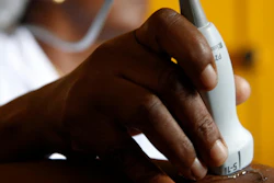
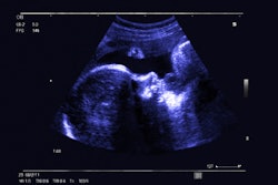
Stringer and colleagues developed a deep-learning AI model to estimate gestational age from blind ultrasonography sweeps. They also incorporated the model into the software of a low-cost, battery-powered device (Butterfly IQ+, Butterfly Network). The study sought to determine the diagnostic accuracy of the device when in the hands of inexperienced users.
The team’s prospective study included 400 women with viable, single, nonanomalous, first-trimester pregnancies in Lusaka, Zambia, and Chapel Hill, NC. For the study, expert sonographers established the ground-truth gestational age through transvaginal crown-rump length measurement.
The novice users meanwhile attended a one-day training session on using the AI ultrasound device and were blinded to the established ground truth gestational age. The same went for the experts.
At random follow-up visits throughout gestation, including a primary evaluation window from 14- to 27-weeks’ gestation, the novice users gathered blind sweeps of the maternal abdomen using the AI-enabled device (index test). The experts meanwhile performed fetal biometry with a high-specification machine (study standard).
In the primary evaluation window, the AI-enabled device performed as well as the study standard. This included a mean absolute error of 3.2 days versus 3 days.
The researchers also observed that the percentage of assessments within seven days of the ground truth gestational age was comparable. This included 90.7% for the index test compared to 92.5% for the study standard.
Finally, the team reported consistent performance in prespecified subgroups, including the Zambia and North Carolina cohorts and women with high body mass index (BMI).
The study authors highlighted that with these results in mind, the portable AI device can be used in low-resource settings. They added that this aligns with the World Health Organization’s (WHO) goal of ultrasonography estimation of gestational age for all pregnant people.
“We hope this can lead to better management of pregnancies, timely interventions, and improved maternal and neonatal health outcomes,” Stringer said.
He told AuntMinnie.com that the research team has a list of additional diagnoses that the technology can make, including diagnosing twins, assessing amniotic fluid volume, and diagnosing malpresentation.
“And we are working on others,” he added. “Our goal is to create a suite of tools that allow the entire spectrum of obstetric sonography to be extended to the most resource-constrained areas of the world.”
In an accompanying editorial, a team led by Alexis Gimovsky, MD, from the Women & Infants Hospital of Rhode Island, wrote that while AI has the potential to advance obstetric care and imaging, techniques such as the one used by Stringer et al are “far from a silver bullet.”
They opined that ultrasonography, whether standard or AI-powered, will only improve such care for pregnant women and newborns when it is implemented within a broader healthcare system in low- and middle-income countries. This includes trained healthcare professionals, basic medications and equipment, and a referral system for managing pregnancy complications.
“Addressing these infrastructure and clinical care challenges is essential for maximizing the impact of advanced ultrasonography technologies in improving maternal and child health outcomes globally,” Gimovsky and co-authors wrote.
The study was funded by the Bill & Melinda Gates Foundation. The full results can be found here.