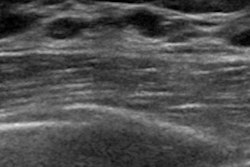
Careful consideration of results from coronal reconstruction on automated breast ultrasound (ABUS) can help clinicians avoid false negatives, according to study results shared February 29 at ECR 2024.
The fact that ABUS is capable of this 3D technique differentiates it from handheld ultrasound, said presenter Elizabet Nikolova, MD, of University Hospital of Zürich in Switzerland. In a study she conducted with colleagues, two women were diagnosed with invasive carcinoma on follow-up after initial ultrasound findings were classified as BI-RADS 2.
"Both lesions were already visible in previous ABUS and mammography and the 'retraction phenomenon sign' [which is highly specific for distinguishing benign from malignant breast lesions] was visible in the ABUS-coronal reconstruction," she said.
Compared with handheld ultrasound, ABUS reduces operator dependence and has a higher reproducibility. And like handheld breast ultrasound, ABUS increases the cancer detection rate when it's used as an adjunct to mammography in women with dense breast tissue, Nikolova noted.
But ABUS does have a few limiting factors that are barriers to its use, she explained, including users' fears of missing lesions, due to artifacts (which can lead to false negatives), incorrect interpretation of findings (which can translate to false positives), and higher cost.
Nikolova and colleagues investigated the false-positive and false-negative rates for ABUS over three years of using the technology in their academic radiology department. They conducted a study that included 1,219 women who underwent ABUS between October 2015 and October 2018. Study participants met the following criteria: all received follow-up at least 24 months after their initial ABUS exam; all were categorized as BI-RADS 4 or 5; and all had histological evaluation. The team defined false-positive cases as those with lesions classified as BI-RADS 3, 4, or 5 in ABUS which were determined to be benign during follow-up or after biopsy, and defined false-negative cases as all those for which cancer was diagnosed during follow-up (and that had lesions retrospectively visible in the initial ABUS examination).
The study participants were classified after ABUS as follows:
- BI-RADS 1: 136, or 11.1%
- BI-RADS 2: 890, or 73%
- BI-RADS 3: 161, or 13.2% (of these, 3, or 1.9%, were identified as malignant)
- BI-RADS 4: 15, or 1.2%, (of these, 7, or 46.6%, were identified as malignant)
- BI-RADS 5: 17, or 1.4% (of these, 100% were identified as malignant)
Of the 1,219 cases, 29 were malignant, for a rate of 2.9%. Two of these cancers were only identified on ABUS. Overall, the study found a false-negative rate of 6.9% and a false-positive rate of 13.9%.
The false negatives prompted Nikolova's group to urge their peers to pay close attention to ABUS coronal reconstruction.
"To avoid false negative cases, ABUS coronal reconstruction should be accurately evaluated," she concluded.
For more coverage from ECR 2024, please visit our RADCast.