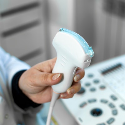
Ultrasound guidance helps shorten cannulation times for extracorporeal cardiopulmonary resuscitation (CPR), according to findings published August 6 in Resuscitation.
Researchers led by Keita Nakatsutsumi from the Tokyo Medical and Dental University Hospital in Japan found that ultrasound guidance led to a decrease in cannulation time by 2.5 minutes compared with conventional cannulation techniques.
"This result should contribute to the development of standard protocols for cannulation in extracorporeal CPR, with the goal of improving outcomes," the team wrote.
Extracorporeal CPR helps bring cardiac arrest patients to treatment, but this requires much technical expertise to apply percutaneous venoarterial extracorporeal membrane oxygenation. Ultrasound guidance is typically used for vascular access, but the researchers pointed out that its effects on cannulation time on these patients are "poorly defined."
Nakatsutsumi and co-authors investigated the effects of real-time ultrasound-guided cannulation for extracorporeal CPR. They used multicenter data from 1,451 cases, of which 510 used ultrasound guidance and 941 were in the control group. The team also evaluated 443 propensity score-matched pairs from the cases.
The researchers found that before matching, cannulation time for ultrasound guidance was 11 minutes, 13 seconds. After matching, the average time was about 11 minutes, while the average time for the control group was about 14 minutes. The researchers reported that these differences were significant in both matched (p < 0.001) and unmatched groups (p = 0.018).
The team also found no significant differences in the incidence of catheter-related complications and incidence of poor neurological outcomes in both groups.
The study authors suggested that, based on their findings and those of previous studies, shortening cannulation time by "even a few minutes" could help improve outcomes for patients with cardiac arrest.
While the team's results did not show significant associations between ultrasound guidance and better survival or neurological outcomes, the authors wrote that "there might be a cohort that could benefit from the time-saving effect of real-time ultrasound-guided cannulation."
Still, the authors called for more studies to determine which factors, in addition to short cannulation time, promote improved neurological outcomes in patients who receive extracorporeal CPR.
The study can be found in its entirety here.


![Examples of ultrasound findings and techniques. (A) Images in a 39-year-old male patient with a mass in the left thigh. The mass is heterogeneous on the B-mode US image (compared with the patient in D) and showed increased microvascularity (superb microvascular imaging [SMI]) and shear-wave elastography (SWE) values. Undifferentiated pleomorphic sarcoma was diagnosed at biopsy (with pleomorphic rhabdomyosarcoma in surgical specimen). (B) Images in an 18-year-old male patient with a mass in the left leg. The mass is hypoechoic on the B-mode image, with no other findings suggestive of malignancy. The lesion is in contact with the cortex of the tibia, which is slightly irregular. CT revealed a doubtful anteromedial tibial erosion. The microvascular study demonstrated high vascularization, suggestive of malignancy. Periosteal Ewing sarcoma was diagnosed with both histologic and immunohistochemical confirmation. (C) Images in a 69-year-old female patient with a lump growing on the outside of the left leg. Multiple SWE examinations were performed (please note the high values obtained in the measurements, whereas the color map highlights the stiffness relative to adjacent tissues). SMI showed areas of increased vascularization to target for sampling. Undifferentiated spindle cell sarcoma was diagnosed at biopsy, with residual leiomyosarcoma in the surgical specimen after neoadjuvant therapy. (D) Images in a 56-year-old female patient with a mass in the right thigh. The mass is heterogeneous at both B-mode ultrasound (similar to patient A) and MRI (coronal T2-weighted spectral attenuated inversion recovery [SPAIR]; T1-weighted pre-contrast and postcontrast imaging), which even shows uptake after the administration of paramagnetic contrast material, which is traditionally suggestive of malignancy. Low values at SMI and elastography are suggestive of benignity. Spindle cell lipoma was diagnosed at biopsy, with atypical spindle cell lipomatous tumor in the surgical specimen.](https://img.auntminnie.com/mindful/smg/workspaces/default/uploads/2026/08/images-radiol250278fig2.APCFLSvX6p.jpg?auto=format%2Ccompress&fit=crop&h=100&q=70&w=100)


![Examples of ultrasound findings and techniques. (A) Images in a 39-year-old male patient with a mass in the left thigh. The mass is heterogeneous on the B-mode US image (compared with the patient in D) and showed increased microvascularity (superb microvascular imaging [SMI]) and shear-wave elastography (SWE) values. Undifferentiated pleomorphic sarcoma was diagnosed at biopsy (with pleomorphic rhabdomyosarcoma in surgical specimen). (B) Images in an 18-year-old male patient with a mass in the left leg. The mass is hypoechoic on the B-mode image, with no other findings suggestive of malignancy. The lesion is in contact with the cortex of the tibia, which is slightly irregular. CT revealed a doubtful anteromedial tibial erosion. The microvascular study demonstrated high vascularization, suggestive of malignancy. Periosteal Ewing sarcoma was diagnosed with both histologic and immunohistochemical confirmation. (C) Images in a 69-year-old female patient with a lump growing on the outside of the left leg. Multiple SWE examinations were performed (please note the high values obtained in the measurements, whereas the color map highlights the stiffness relative to adjacent tissues). SMI showed areas of increased vascularization to target for sampling. Undifferentiated spindle cell sarcoma was diagnosed at biopsy, with residual leiomyosarcoma in the surgical specimen after neoadjuvant therapy. (D) Images in a 56-year-old female patient with a mass in the right thigh. The mass is heterogeneous at both B-mode ultrasound (similar to patient A) and MRI (coronal T2-weighted spectral attenuated inversion recovery [SPAIR]; T1-weighted pre-contrast and postcontrast imaging), which even shows uptake after the administration of paramagnetic contrast material, which is traditionally suggestive of malignancy. Low values at SMI and elastography are suggestive of benignity. Spindle cell lipoma was diagnosed at biopsy, with atypical spindle cell lipomatous tumor in the surgical specimen.](https://img.auntminnie.com/mindful/smg/workspaces/default/uploads/2026/08/images-radiol250278fig2.APCFLSvX6p.jpg?auto=format%2Ccompress&fit=crop&h=112&q=70&w=112)