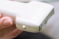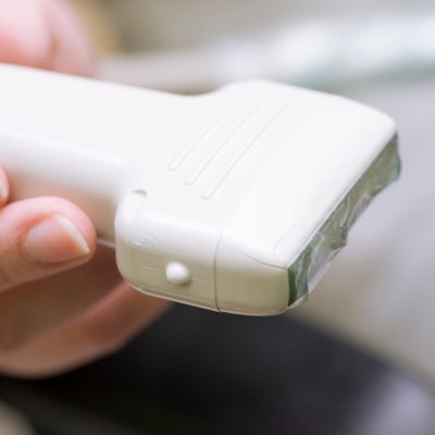
Point-of-care ultrasound (POCUS) can go toe-to-toe with traditional, cart-based ultrasound systems when it comes to studying multiple organs, according to findings presented March 29 at UltraCon.
Dr. Ryan Gibbons from Temple University in Media, PA, discussed his team's study, which found that POCUS has similar diagnostic accuracy compared to cart systems. The group tested this on cardiac, lung, renal, aorta, and gallbladder ultrasound studies.
"I think the fact that these handheld devices seem to be as good as a traditional cart-based model at a fraction of the cost will really help move point-of-care [ultrasound] forward," Gibbons told AuntMinnie.com.
Previous studies have highlighted POCUS' potential in diagnosing pathologies. Gibbons said these portable, handheld devices have helped expand the clinical scope of POCUS while being less costly than traditional cart-based ultrasound models.
"At our institution, we've been incorporating [POCUS devices] into medical school teaching, and also in the hospital," Gibbons said. "Our students have their own handheld devices."
However, he also noted that there is a lack of data assessing the diagnostic accuracy of POCUS devices. So Gibbons and colleagues conducted a study to compare the diagnostic accuracy of POCUS versus traditional cart-based ultrasound.
The study included 118 patients, 61 of whom underwent a cart-based ultrasound exam and 57 of whom were scanned with the handheld device. Of the total number of exams, 32 were cardiac, 23 lung, 26 renal, 15 aorta, and 22 gallbladder.
Each POCUS exam was performed by either an ultrasound fellow or a year one-to-three emergency medicine resident. Reference standards for these readings came from a cardiologist-interpreted echocardiogram, a radiology-interpretated ultrasound or CT exam, or POCUS quality review by three ultrasound fellowship-trained physicians.
Gibbons and colleagues found that the diagnostic accuracy of the handheld device was 91.1% (range, 80.4% to 97%) compared with 85% seen for the cart-based model (p = 0.315).
Gibbons did note that cart-based ultrasound had better image quality. Nevertheless, he emphasized that the results show how POCUS can be used in educational and clinical settings and that the technology is "well on its way" to being more widely used.
He also told AuntMinnie.com that his team plans to further investigate the performance of POCUS for a variety of exams.
"We were kind of broad in what we did," he said. "We looked at five different pathologies. We need to look at them individually and conduct a larger study for each to support what we found in this initial study."