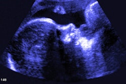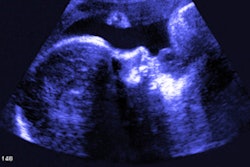
Ultrasound should be used to evaluate women who present with vaginal bleeding symptoms after embryo transfer via in vitro fertilization (IVF), suggest findings published July 23 in the American Journal of Obstetrics and Gynecology.
Researchers led by Meredith Pollie, MD, from Cornell University in New York found that combining ultrasound measurements with early pregnancy bleeding can help better assess spontaneous abortion risk among women with a history of infertility.
"In the setting of normal ultrasound measurements, patients can be reassured that their risk for spontaneous abortion is not increased, and risk for live birth is not decreased," Pollie and colleagues wrote.
Previous reports suggest that while about 15% of clinically recognized pregnancies in infertile women end via spontaneous abortion, it's not well described in women's natural history. Vaginal bleeding in the early stages of pregnancy is a common symptom in these women who have turned to methods such as embryo transfers from IVF. However, the researchers noted that the prognostic value of this symptom isn't well known.
Pollie and colleagues wanted to investigate how combining this symptom with first-trimester ultrasound measurements could potentially measure spontaneous abortion risk.
They included retrospective data from 1,858 women with singleton pregnancies between 2019 and 2019. Of these, 359 had spontaneous abortions and 1,499 had live births. Also, of the total, 17% (n = 315) reported vaginal bleeding.
The team found significant ties between combined ultrasound measurements and bleeding with spontaneous abortion at six weeks' gestation, but only when accompanied by absent fetal heart rate or both absent fetal heart rate and absent fetal pole. This included relative risk (RR) values of 5.36 and 9.67, respectively.
Additionally, the researchers found similar trends at seven weeks' gestation, though higher RR values were found only when accompanied by abnormal assessment of fetal heart rate or crown rump length (RR = 5.09) or both fetal heart rate and crown rump length (RR = 14.82).
Finally, they found that with normal ultrasound measurements, bleeding had no significant associations with increased spontaneous abortion risk at six weeks' gestation (RR = 1.05) and seven weeks' gestation (RR = 0.8). They reported that the live birth rate was approximate to women with normal imaging measurements and no vaginal bleeding present.
The study authors suggested that based on their results, expecting mothers who present with vaginal bleeding but with normal ultrasound measurements can be reassured of their pregnancies. They added that their data can be used to identify and guide counseling for women with abnormal imaging measurements who may be at higher risk for spontaneous abortion.
The study can be found in its entirety here.


![Examples of ultrasound findings and techniques. (A) Images in a 39-year-old male patient with a mass in the left thigh. The mass is heterogeneous on the B-mode US image (compared with the patient in D) and showed increased microvascularity (superb microvascular imaging [SMI]) and shear-wave elastography (SWE) values. Undifferentiated pleomorphic sarcoma was diagnosed at biopsy (with pleomorphic rhabdomyosarcoma in surgical specimen). (B) Images in an 18-year-old male patient with a mass in the left leg. The mass is hypoechoic on the B-mode image, with no other findings suggestive of malignancy. The lesion is in contact with the cortex of the tibia, which is slightly irregular. CT revealed a doubtful anteromedial tibial erosion. The microvascular study demonstrated high vascularization, suggestive of malignancy. Periosteal Ewing sarcoma was diagnosed with both histologic and immunohistochemical confirmation. (C) Images in a 69-year-old female patient with a lump growing on the outside of the left leg. Multiple SWE examinations were performed (please note the high values obtained in the measurements, whereas the color map highlights the stiffness relative to adjacent tissues). SMI showed areas of increased vascularization to target for sampling. Undifferentiated spindle cell sarcoma was diagnosed at biopsy, with residual leiomyosarcoma in the surgical specimen after neoadjuvant therapy. (D) Images in a 56-year-old female patient with a mass in the right thigh. The mass is heterogeneous at both B-mode ultrasound (similar to patient A) and MRI (coronal T2-weighted spectral attenuated inversion recovery [SPAIR]; T1-weighted pre-contrast and postcontrast imaging), which even shows uptake after the administration of paramagnetic contrast material, which is traditionally suggestive of malignancy. Low values at SMI and elastography are suggestive of benignity. Spindle cell lipoma was diagnosed at biopsy, with atypical spindle cell lipomatous tumor in the surgical specimen.](https://img.auntminnie.com/mindful/smg/workspaces/default/uploads/2026/08/images-radiol250278fig2.APCFLSvX6p.jpg?auto=format%2Ccompress&fit=crop&h=100&q=70&w=100)


![Examples of ultrasound findings and techniques. (A) Images in a 39-year-old male patient with a mass in the left thigh. The mass is heterogeneous on the B-mode US image (compared with the patient in D) and showed increased microvascularity (superb microvascular imaging [SMI]) and shear-wave elastography (SWE) values. Undifferentiated pleomorphic sarcoma was diagnosed at biopsy (with pleomorphic rhabdomyosarcoma in surgical specimen). (B) Images in an 18-year-old male patient with a mass in the left leg. The mass is hypoechoic on the B-mode image, with no other findings suggestive of malignancy. The lesion is in contact with the cortex of the tibia, which is slightly irregular. CT revealed a doubtful anteromedial tibial erosion. The microvascular study demonstrated high vascularization, suggestive of malignancy. Periosteal Ewing sarcoma was diagnosed with both histologic and immunohistochemical confirmation. (C) Images in a 69-year-old female patient with a lump growing on the outside of the left leg. Multiple SWE examinations were performed (please note the high values obtained in the measurements, whereas the color map highlights the stiffness relative to adjacent tissues). SMI showed areas of increased vascularization to target for sampling. Undifferentiated spindle cell sarcoma was diagnosed at biopsy, with residual leiomyosarcoma in the surgical specimen after neoadjuvant therapy. (D) Images in a 56-year-old female patient with a mass in the right thigh. The mass is heterogeneous at both B-mode ultrasound (similar to patient A) and MRI (coronal T2-weighted spectral attenuated inversion recovery [SPAIR]; T1-weighted pre-contrast and postcontrast imaging), which even shows uptake after the administration of paramagnetic contrast material, which is traditionally suggestive of malignancy. Low values at SMI and elastography are suggestive of benignity. Spindle cell lipoma was diagnosed at biopsy, with atypical spindle cell lipomatous tumor in the surgical specimen.](https://img.auntminnie.com/mindful/smg/workspaces/default/uploads/2026/08/images-radiol250278fig2.APCFLSvX6p.jpg?auto=format%2Ccompress&fit=crop&h=112&q=70&w=112)