
Measures from thyroid nodule vascularity and shear-wave elastography show promise for improving the detection of thyroid cancer, according to findings presented at the American Roentgen Ray Society (ARRS) annual meeting in Honolulu.
Researchers led by Dr. Leila Aghaghazvini from the Iran University of Medical Sciences in Tehran found that using these measures adds diagnostic value to traditional TI-RADS assessment of suspicious thyroid nodules, including higher sensitivity and specificity among other achievement marks.
"It would be advantageous to incorporate these criteria into TI-RADS risk stratification systems in order to reduce unnecessary fine-needle aspirations while keeping a high detection sensitivity for malignant tumors," Aghaghazvini and colleagues wrote.
Conventional ultrasound is typically used to detect thyroid cancer after physical examination and palpitation. However, Aghaghazvini et al noted that there is a lack of evidence that any particular ultrasound findings can predict thyroid cancer.
Specialists use TI-RADS to classify risk of malignancy based on ultrasound findings. But Aghaghazvini said that this does take a nodule's vascular or elastic characteristics into account. To address this knowledge gap, the team explored the value of adding vascularity and shear-wave elastography assessments to conventional TI-RADS assessments.
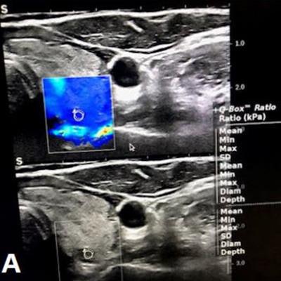
 In an online poster presentation at the ARRS annual meeting, researchers led by Dr. Leila Aghaghazvini found that elastography and vascularity measures can help improve the performance of TI-RADS in assessing suspicious thyroid nodules. In these images, gray-scale features show a markedly hypoechoic thyroid nodule with irregular margins (TI-RADS 4), later confirmed as papillary thyroid cancer. (A) Elastography shows a mostly blue pattern. (B) Doppler sonography did not show any flow pattern. Images courtesy of the ARRS.
The study included 200 total thyroid nodules, assessed by conventional sonography to gather TI-RADS scores and important characteristics. The investigators used a 7.5 MHz probe to assess vascularity pattern and resistive index, performed quantitative elastography evaluations via color mapping, and calculated average and maximum velocities. They confirmed final diagnoses of all thyroid nodules via histopathology assessment or follow-up imaging.
The study found 37 TI-RADS 4 nodules, 15 of which were malignant, and 57 TI-RADS 5 nodules, 12 of which were malignant.
Overall, area under the curve (AUC) for TI-RADS assessment was 0.76. Adding elastography and resistive index measures to the mix translated to higher AUC values for TI-RADS 4 and 5 nodules.
| AUC values of vascularity and elastography measures | ||
| Measure | TI-RADS 4 | TI-RADS 5 |
| Resistive index | 0.93 | 0.96 |
| Color map elastography grades | 0.89 | 0.85 |
| Shear-wave elastography maximum velocity | 0.86 | 0.96 |
| Shear-wave elastography average velocity | 0.82 | 0.97 |
| Doppler grade | 0.8 | 0.8 |
The researchers also used a resistive index cutoff point of 0.6 in TI-RADS 4 nodules and found even higher values. This included AUCs of 0.93 for sensitivity, 1 for specificity, 0.97 for efficiency, 1 for positive predictive value, and 0.96 for negative predictive value.
When a cutoff point of 4.33 was used for shear-wave elastography average velocity, the researchers observed high diagnostic efficacy values. These included AUCs of 1 for sensitivity, 0.82 for specificity, 0.86 for efficiency, 0.6 for positive predictive value, and 1 for negative predictive value.
The study authors hope that these assessments will improve differentiation between malignant and benign thyroid nodules, Aghaghazvini said.
![Examples of ultrasound findings and techniques. (A) Images in a 39-year-old male patient with a mass in the left thigh. The mass is heterogeneous on the B-mode US image (compared with the patient in D) and showed increased microvascularity (superb microvascular imaging [SMI]) and shear-wave elastography (SWE) values. Undifferentiated pleomorphic sarcoma was diagnosed at biopsy (with pleomorphic rhabdomyosarcoma in surgical specimen). (B) Images in an 18-year-old male patient with a mass in the left leg. The mass is hypoechoic on the B-mode image, with no other findings suggestive of malignancy. The lesion is in contact with the cortex of the tibia, which is slightly irregular. CT revealed a doubtful anteromedial tibial erosion. The microvascular study demonstrated high vascularization, suggestive of malignancy. Periosteal Ewing sarcoma was diagnosed with both histologic and immunohistochemical confirmation. (C) Images in a 69-year-old female patient with a lump growing on the outside of the left leg. Multiple SWE examinations were performed (please note the high values obtained in the measurements, whereas the color map highlights the stiffness relative to adjacent tissues). SMI showed areas of increased vascularization to target for sampling. Undifferentiated spindle cell sarcoma was diagnosed at biopsy, with residual leiomyosarcoma in the surgical specimen after neoadjuvant therapy. (D) Images in a 56-year-old female patient with a mass in the right thigh. The mass is heterogeneous at both B-mode ultrasound (similar to patient A) and MRI (coronal T2-weighted spectral attenuated inversion recovery [SPAIR]; T1-weighted pre-contrast and postcontrast imaging), which even shows uptake after the administration of paramagnetic contrast material, which is traditionally suggestive of malignancy. Low values at SMI and elastography are suggestive of benignity. Spindle cell lipoma was diagnosed at biopsy, with atypical spindle cell lipomatous tumor in the surgical specimen.](https://img.auntminnie.com/mindful/smg/workspaces/default/uploads/2026/08/images-radiol250278fig2.APCFLSvX6p.jpg?auto=format%2Ccompress&fit=crop&h=100&q=70&w=100)



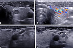
![Examples of ultrasound findings and techniques. (A) Images in a 39-year-old male patient with a mass in the left thigh. The mass is heterogeneous on the B-mode US image (compared with the patient in D) and showed increased microvascularity (superb microvascular imaging [SMI]) and shear-wave elastography (SWE) values. Undifferentiated pleomorphic sarcoma was diagnosed at biopsy (with pleomorphic rhabdomyosarcoma in surgical specimen). (B) Images in an 18-year-old male patient with a mass in the left leg. The mass is hypoechoic on the B-mode image, with no other findings suggestive of malignancy. The lesion is in contact with the cortex of the tibia, which is slightly irregular. CT revealed a doubtful anteromedial tibial erosion. The microvascular study demonstrated high vascularization, suggestive of malignancy. Periosteal Ewing sarcoma was diagnosed with both histologic and immunohistochemical confirmation. (C) Images in a 69-year-old female patient with a lump growing on the outside of the left leg. Multiple SWE examinations were performed (please note the high values obtained in the measurements, whereas the color map highlights the stiffness relative to adjacent tissues). SMI showed areas of increased vascularization to target for sampling. Undifferentiated spindle cell sarcoma was diagnosed at biopsy, with residual leiomyosarcoma in the surgical specimen after neoadjuvant therapy. (D) Images in a 56-year-old female patient with a mass in the right thigh. The mass is heterogeneous at both B-mode ultrasound (similar to patient A) and MRI (coronal T2-weighted spectral attenuated inversion recovery [SPAIR]; T1-weighted pre-contrast and postcontrast imaging), which even shows uptake after the administration of paramagnetic contrast material, which is traditionally suggestive of malignancy. Low values at SMI and elastography are suggestive of benignity. Spindle cell lipoma was diagnosed at biopsy, with atypical spindle cell lipomatous tumor in the surgical specimen.](https://img.auntminnie.com/mindful/smg/workspaces/default/uploads/2026/08/images-radiol250278fig2.APCFLSvX6p.jpg?auto=format%2Ccompress&dpr=2&fit=crop&h=167&q=70&w=250)


