Renal denervation via ultrasound can reduce daytime ambulatory systolic blood pressure when antihypertensive medications aren't available, suggest findings from a French study published February 28 in JAMA.
A team led by Dr. Michel Azizi, PhD, from European Hospital Georges Pompidou in Paris found that compared with a sham procedure without postprocedural major adverse events, renal ultrasound denervation showed a reduction in daytime ambulatory systolic blood pressure in hypertension patients at two months.
"These results solidify the role of the ultrasound renal denervation system as an adjunctive therapy for hypertension treatment, in addition to medications and lifestyle modification," Azizi told AuntMinnie.com.
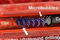
Hypertension is tricky to treat, and its prevalence is rising, previous reports suggest. Previous sham-controlled trials looked at the efficacy of ultrasound renal denervation and radiofrequency-based renal denervation for lowering blood pressure. These trials used a catheter-based system using ultrasound renal denervation to achieve consistent ablation of renal sympathetic nerves.
Azizi and colleagues designed their trial, called Radiance II, to further the study these previous trials set out to accomplish. They wanted to explore the potential efficacy and safety of the ultrasound renal denervation procedure in the absence of antihypertensive medications.
In this sham-controlled, randomized clinical trial, patients between the ages of 18 and 75 with hypertension underwent either ultrasound renal denervation or a sham procedure. Patients were to abstain from antihypertensive medications until the two-month follow-up.
The researchers included data from a sample of 224 patients, including 150 who were randomized to denervation and 74 who underwent a sham procedure. They found at two months that the reduction in daytime ambulatory systolic blood pressure was greater in the ultrasound renal denervation group. This included an average reduction of 7.9 mm of mercury (mmHg), compared with a 1.8 mmHg reduction in the sham group (p < 0.001).
The team also noted a "consistent" effect of ultrasound renal denervation throughout the 24-hour circadian cycle. Among seven secondary blood pressure outcomes, the group found that six were significantly improved with ultrasound renal denervation versus the sham procedure. Finally, no major adverse events were reported in either group.
Azizi added that these results are in line with a consensus statement made in February by the European Society of Cardiology's Council on Hypertension and the European Association of Percutaneous Cardiovascular Interventions for renal denervation in hypertension. He said that pooled analysis showed that denervation can safely lower blood pressure in patients with varying degrees of hypertension severity.
The study authors wrote that while their trial was paused and then restarted due to the COVID-19 pandemic, they found that enrollment prior to or after the pandemic did not influence the blood pressure response to ultrasound renal denervation.
The authors also noted that the long-term safety of ultrasound renal denervation will continue to be monitored for 60 months in the current trial with renal CT angiography or MR angiography imaging at 12 months in the denervation group.
Azizi also told AuntMinnie.com that the team is looking for short- and long-term predictors of blood pressure response to help physicians to take decisions to propose ultrasound renal denervation to patients.