Tuesday, November 29 | 3:00 p.m.-4:00 p.m. | T7-SSBR07-5 | E450B
In this presentation, research will show how using ultrasound first to assess noncalcified lesions recalled by digital breast tomosynthesis (DBT) is cost-effective and can improve patient quality of life.Dr. Berat Bersu Ozcan from the University of Texas Southwestern Medical Center in Dallas will discuss her team's findings, which compared the economic feasibility of using ultrasound versus digital mammography in this setting. Specifically, the team looked at overall cost and the impact of each outcome in quality-adjusted life years (QALY). They calculated cost-effectiveness by incremental cost-effectiveness ratios (ICER) and net monetary benefit (NMB) calculations.
The researchers found that using ultrasound first gave a total cost of $462 and 0.986 QALYs compared with $446 and 0.983 QALYs for digital mammography first. They also found that NMB was $98,130 for ultrasound first, compared with $97,857 for using digital mammography first. Additionally, an ICER of $5,546.65/QALY gained was found for using ultrasound first over mammography, lower than the willingness-to-pay threshold of $100,000 per QALY.
"Ultrasound first was the better strategy in 70% of the iterations according to the probabilistic sensitivity analysis," the study authors wrote.
Based on their results, the researchers suggested that ultrasound is more economically feasible than using mammography first when analyzing DBT-recalled noncalcified lesions. Find out more in this session.




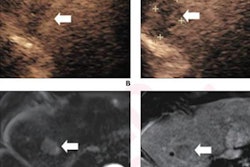



![Examples of ultrasound findings and techniques. (A) Images in a 39-year-old male patient with a mass in the left thigh. The mass is heterogeneous on the B-mode US image (compared with the patient in D) and showed increased microvascularity (superb microvascular imaging [SMI]) and shear-wave elastography (SWE) values. Undifferentiated pleomorphic sarcoma was diagnosed at biopsy (with pleomorphic rhabdomyosarcoma in surgical specimen). (B) Images in an 18-year-old male patient with a mass in the left leg. The mass is hypoechoic on the B-mode image, with no other findings suggestive of malignancy. The lesion is in contact with the cortex of the tibia, which is slightly irregular. CT revealed a doubtful anteromedial tibial erosion. The microvascular study demonstrated high vascularization, suggestive of malignancy. Periosteal Ewing sarcoma was diagnosed with both histologic and immunohistochemical confirmation. (C) Images in a 69-year-old female patient with a lump growing on the outside of the left leg. Multiple SWE examinations were performed (please note the high values obtained in the measurements, whereas the color map highlights the stiffness relative to adjacent tissues). SMI showed areas of increased vascularization to target for sampling. Undifferentiated spindle cell sarcoma was diagnosed at biopsy, with residual leiomyosarcoma in the surgical specimen after neoadjuvant therapy. (D) Images in a 56-year-old female patient with a mass in the right thigh. The mass is heterogeneous at both B-mode ultrasound (similar to patient A) and MRI (coronal T2-weighted spectral attenuated inversion recovery [SPAIR]; T1-weighted pre-contrast and postcontrast imaging), which even shows uptake after the administration of paramagnetic contrast material, which is traditionally suggestive of malignancy. Low values at SMI and elastography are suggestive of benignity. Spindle cell lipoma was diagnosed at biopsy, with atypical spindle cell lipomatous tumor in the surgical specimen.](https://img.auntminnie.com/mindful/smg/workspaces/default/uploads/2026/08/images-radiol250278fig2.APCFLSvX6p.jpg?auto=format%2Ccompress&fit=crop&h=112&q=70&w=112)


