Dear AuntMinnie Member,
Is there anything artificial intelligence (AI) can't do? Our top story for the past week is coverage of a fascinating study in which researchers from China developed an AI algorithm that creates x-ray-like images from ultrasound scans.
Why not just use x-ray? The researchers pointed out that the AI-generated images could be useful for some patients who are particularly sensitive to radiation, such as children who might need to be monitored on an ongoing basis for scoliosis progression.
Find out how the algorithm works in an article in our Ultrasound Community. And while you're there, check out our coverage of the latest research on the following:
- A homegrown ultrasound phantom cooked up from ingredients you would find in your kitchen
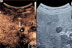
- Liver biopsy guided by contrast-enhanced ultrasound is more effective than guidance under conventional ultrasound
- A prediction model that uses contrast ultrasound can help predict prostate cancer risk in men
PET for Alzheimer's
Meanwhile, researchers from Canada used PET with a combination of radiopharmaceuticals to assess beta amyloid and tau markers in people who might be at risk of progressing to long-term cognitive decline -- before they even show signs of cognitive impairment.
They found that PET scans were able to identify which individuals were at risk of long-term cognitive decline that could progress to Alzheimer's disease. Learn more in an article in our Molecular Imaging Community.
AI webinar on Aug. 10
Finally, have you registered yet for our free webinar on getting paid for AI in radiology, scheduled for 4 p.m. Eastern Time on Wednesday, August 10? If not, we still have seats available ... as always, registration is free.







![Examples of ultrasound findings and techniques. (A) Images in a 39-year-old male patient with a mass in the left thigh. The mass is heterogeneous on the B-mode US image (compared with the patient in D) and showed increased microvascularity (superb microvascular imaging [SMI]) and shear-wave elastography (SWE) values. Undifferentiated pleomorphic sarcoma was diagnosed at biopsy (with pleomorphic rhabdomyosarcoma in surgical specimen). (B) Images in an 18-year-old male patient with a mass in the left leg. The mass is hypoechoic on the B-mode image, with no other findings suggestive of malignancy. The lesion is in contact with the cortex of the tibia, which is slightly irregular. CT revealed a doubtful anteromedial tibial erosion. The microvascular study demonstrated high vascularization, suggestive of malignancy. Periosteal Ewing sarcoma was diagnosed with both histologic and immunohistochemical confirmation. (C) Images in a 69-year-old female patient with a lump growing on the outside of the left leg. Multiple SWE examinations were performed (please note the high values obtained in the measurements, whereas the color map highlights the stiffness relative to adjacent tissues). SMI showed areas of increased vascularization to target for sampling. Undifferentiated spindle cell sarcoma was diagnosed at biopsy, with residual leiomyosarcoma in the surgical specimen after neoadjuvant therapy. (D) Images in a 56-year-old female patient with a mass in the right thigh. The mass is heterogeneous at both B-mode ultrasound (similar to patient A) and MRI (coronal T2-weighted spectral attenuated inversion recovery [SPAIR]; T1-weighted pre-contrast and postcontrast imaging), which even shows uptake after the administration of paramagnetic contrast material, which is traditionally suggestive of malignancy. Low values at SMI and elastography are suggestive of benignity. Spindle cell lipoma was diagnosed at biopsy, with atypical spindle cell lipomatous tumor in the surgical specimen.](https://img.auntminnie.com/mindful/smg/workspaces/default/uploads/2026/08/images-radiol250278fig2.APCFLSvX6p.jpg?auto=format%2Ccompress&fit=crop&h=112&q=70&w=112)


