Point-of-care ultrasound (POCUS) for lung imaging is noninferior to chest x-ray when it comes to diagnosing COVID-19, according to an Italian study published January 18 in the Journal of Ultrasound in Medicine.
A team led by Dr. Costantino Caroselli from the National Institute of Rest and Care for the Elderly in Ancona found that ultrasound characteristics seen in COVID-positive patients included B-lines, irregular pleural lines, and small consolidation.
"Lung ultrasound holds the promise of an accurate, radiation-free, and affordable diagnostic and monitoring tool in COVID-19 pneumonia," Caroselli and colleagues wrote.
Lung imaging is an important tool in helping diagnose and manage COVID-19 in patients. Previous studies have shown ultrasound's promise in determining early lung involvement in patients while preventing radiation exposure.
However, the researchers said that the lack of a unified approach for POCUS among clinicians is keeping this method from wider use. They added this is caused by a lack of data on patient outcomes and lung ultrasound having a lower specificity than chest x-ray.
Caroselli's team wanted to show how useful ultrasound is in diagnosing lung disease early in COVID-positive patients and compare their results to findings on chest x-ray. They looked at data from 479 patients, 82.7% of whom were diagnosed with COVID-19 by molecular testing.
Lung POCUS was performed by trained emergency physicians on patients presenting for possible COVID-19 evaluation before they underwent conventional radiologic examination or while they were waiting for the report.
| Most common findings on lung POCUS in COVID patients | |
| B-lines | 80.2% |
| Irregular pleural lines | 59.3% |
| Small subpleural consolidations | 55.3% |
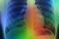
Chest x-rays were found to be normal, meanwhile, in 17.9% of all cases.
The team also tested the prognostic capabilities of lung POCUS. Pleural effusion, small consolidation, male sex, and clinical history were associated with hospital admission rate. However, x-ray outcomes were not associated with this rate.
Both POCUS and x-ray have significant roles in determining the need for orotracheal intubation for patients, the study authors wrote. Lung ultrasound small consolidation, x-ray ground glass, and x-ray interstitial pattern were found to be linked to patients needing invasive mechanical ventilation.
Meanwhile, pleural effusion found on ultrasound and x-ray small consolidation were the only predictors of mortality.
The researchers wrote that lung POCUS could help clinicians rapidly evaluate and triage patients as coronavirus surges persist.
"Our analysis confirmed that, as well as supporting COVID-19 diagnosis, lung ultrasound was useful in prognostic stratification relating to the need for hospitalization, orotracheal intubation, and mortality among the patients studied," they wrote.