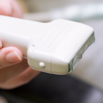
Point-of-care ultrasound (POCUS) is an inclusive technology that isn't limited to one specialty, but questions still need to be answered for more widespread use, according to an article published October 21 in the New England Journal of Medicine.
A team led by Dr. José Díaz-Gómez from the Baylor College of Medicine in Texas found that POCUS provides clinicians with real-time diagnostic and monitoring information and can be used to enhance standard ultrasound-guided procedural safety.
"The introduction of POCUS curricula and training at the medical school and postgraduate levels, the increasing level of evidence of its effect in clinical practice, and advances in handheld systems all point toward the possibility that POCUS will become a standard tool of the frontline clinician," Díaz-Gómez and colleagues wrote.
POCUS has gained in popularity in recent years for its ease of use, portability, and cost-effectiveness. Despite its appeal, POCUS has some unanswered questions surrounding it when it comes to morbidity, mortality, and training.
Some researchers have also called for more rigorously designed trials to prove the value of POCUS. An editorial published in October called for more evidence in clinical benefits and patient outcomes from POCUS.
Díaz-Gómez et al wanted to review previous studies in determining how POCUS should be viewed and used.
The two questions researchers asked in their analysis included the following: "Does POCUS affect patient-centered end points such as functional status, morbidity, and mortality?" and "What specific training is required for a clinician to become competent in POCUS?"
For the first question, the team said it's a challenge to study the effect of POCUS on patient outcomes, given several factors. These include the heterogeneity of patient populations, the difficulty in standardizing scanning protocols, and variation in clinician skill levels, among others.
"It is not likely that focusing on POCUS as the primary variable determining the outcome will be a productive approach to research," the study authors wrote.
For the second question, the researchers called on the need for information in identifying the hours of training needed for mastering image acquisition and interpretation. This includes course design, finding out the usefulness of simulations, and continuing assessment of skill maintenance. They cautioned that careless use of POCUS could lead to health violations.
"A key aspect to the success of a training sequence for POCUS also relates to the skill and motivation of the faculty, particularly with regard to hands-on training. This is an unexamined area of research," they said.
However, the researchers also noted that POCUS is a useful monitoring tool when it comes to assessing cardiac function in patients with COVID-19, as well as heart function before and after cardiopulmonary resuscitation (CPR). They also heralded its comparable accuracy with other imaging methods when it comes to identifying certain disorders, such as abdominal aortic aneurysm and COVID-19 pneumonia.
Research highlighted by the investigators in their analysis also touted POCUS as allowing for fewer return visits and a shorter length of stay compared to conventional approaches.
"However, it will be critical to determine which, if any, [POCUS] applications ultimately benefit patient care," they added.