An artificial intelligence (AI) algorithm designed to interpret compression ultrasound images can distinguish between patients who do and do not have deep vein thrombosis (DVT), according to a study published September 15 in npj Digital Medicine.
What's more, the researchers determined the AI program could save health providers $150 per exam over standard ultrasound methods.
"Traditionally, DVT diagnoses need a specialist ultrasound scan performed by a trained radiographer, and we have found that the preliminary data using the AI algorithm coupled to a hand-held ultrasound machine shows promising results," said senior author Dr. Nicola Curry of Oxford University, London, in a news release.
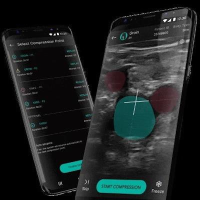
The AI program, called AutoDVT, was developed by ThinkSono.
 ThinkSono AutoDVT App on a smartphone. Image courtesy of ThinkSono.
DVT is a blood clot most commonly found in the leg, and it can lead to fatal pulmonary embolism, according to the study authors. Compression ultrasound of the legs is the diagnostic gold standard for the condition. However, many patients with possible symptoms are found not to have a DVT, which results in long referral waiting times for patients and a large clinical burden for specialists. Thus, diagnosis at the point of care by nonspecialists is desired, the authors wrote.
The researchers trained the AutoDVT program on ultrasound videos from 255 volunteers and evaluated its performance on a sample size of 53 prospectively enrolled patients from a National Health Service DVT diagnostic clinic and 30 prospectively enrolled patients from a German DVT clinic.
The AI DVT program achieved sensitivities between 0.82 to 0.94, specificities of 0.70 to 0.82, and positive predictive values and negative predictive values in ranges of 0.65 to 0.89 and 0.99 to 1.00 when compared with the clinical gold standard, the researchers found.
In addition, the researchers provided a cost analysis for integrating the technology into diagnostic pathways for DVT. They estimated the approach could save health services $150 per exam.
"Currently, many patients do not have a definitive diagnosis within 24 hours of a suspected DVT, and so many patients end up receiving painful injections of what can often be an unnecessary anticoagulant, with potential side-effects," Curry added.
ThinkSono said this is the first study to show that a machine-learning AI algorithm can potentially diagnose DVT and that researchers are due soon to start a clinical study to evaluate the product further.