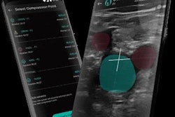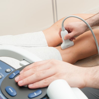
Three different compression ultrasound methods all performed well for imaging deep vein thrombosis (DVT) in a study published online on February 11 by PLOS One. The failure rates for single limited, serial limited, and whole-leg ultrasound ranged from 1% to 2%.
While the three methods are regularly used in clinical practice to help identify DVT, there is no consensus about which one works best. The authors reviewed dozens of studies with thousands of patients and found that no one ultrasound method stood above the rest.
"In the present meta-analysis, more recent studies using clinical follow-up as the reference method showed very comparable failure rates of the three strategies," wrote the authors, led by Dr. Noémie Kraaijpoel from the department of vascular medicine at the University of Amsterdam. "This could imply that there may not be a preferred strategy when taking only safety into account."
The authors searched the scientific literature for compression ultrasound studies from on January 1, 1989, the year the first high-quality research on the modality was published, through July 2019. They included studies using three methods of imaging adults with suspected DVT:
- Single limited compression ultrasound -- a single examination of the proximal deep veins
- Serial limited compression ultrasound -- two examinations about five to 10 days apart of proximal deep veins
- Whole-leg compression ultrasound -- a single examination of both distal and proximal deep veins
Six studies with 2,079 patients assessed single limited compression ultrasound. The analysis showed the modality had a 1.4% failure rate and 6.4% proportion of positive results, although the findings varied among the included individual studies.
For serial limited compression ultrasound, the authors included 11 studies with 3,360 patients. The modality had a 1.9% failure rate and 25% proportion of positive results.
Finally, seven studies with 3,159 evaluated whole-leg compression ultrasound. The findings showed a failure rate of 1% and 25% proportion of positive results.
While all included studies had comparable failure rates (p = 0.36), the proportion of confirmed DVT was significantly higher for serial limited and whole-leg compression ultrasound than for single limited compression ultrasound. The authors noted the difference in prevalence could be the result of patient selection.
Notably, single limited compression ultrasound had a lower DVT prevalence -- likely due to the selection of patients with a lower DVT risk. As a result, the modality could theoretically have a higher failure rate if more DVTs had been found in the group.
Nevertheless, the compression ultrasound methods produced similar results and may be considered equivalent for clinical practice.
"The failure rates of single limited, serial limited, and whole-leg [compression ultrasound] for DVT were found to be quite comparable in patients selected as per the individual study diagnostic algorithms," the authors wrote. "Preference for one of the strategies should be based on probability assessment, feasibility, and expertise."