The market for healthcare artificial intelligence (AI) applications was worth $823 million in 2019, representing the sixth-largest market by revenue for AI. But this is still just a small fraction of the $16 billion overall market for AI -- indicating room for growth, according to market research firm Omdia.
Omdia forecasts that the healthcare AI software market will grow to over $10 billion in 2025. This drastic growth will be driven by the large size of the healthcare equipment market, which totaled $214 billion in 2019, and the great amount of investment available to the healthcare industry.
Despite the great potential provided by the healthcare industry, the healthcare AI market remains mostly untapped, as the technology is still in the early stages of development and faces barriers such as regulation, and many healthcare providers are unsure of the value of AI.
AI vendors have developed advanced AI algorithms that can be applied to healthcare devices, especially for medical imaging. Many major healthcare equipment manufacturers have partnered with these vendors to incorporate AI tools into product lines or have developed AI software tools in-house. Collaborations between healthcare providers, equipment manufacturers, and AI vendors will be crucial to ensuring that healthcare challenges are addressed.
AI has the potential to help combat the growing problem of strained healthcare provision by helping healthcare practices deliver better patient outcomes with limited resources. These challenges have been exacerbated by the pandemic caused by COVID-19, making healthcare AI of the utmost importance.
Healthcare AI, especially for ultrasound, is being developed in collaboration with end users to solve real-world problems. According to the Omdia Artificial Intelligence for Ultrasound Survey (AIUS) 2020, many healthcare practices rely on AI to address areas of improvement, including image quality and standardization, workflow efficiency, and diagnostic support. For example, algorithms assist in prioritizing and screening patients.
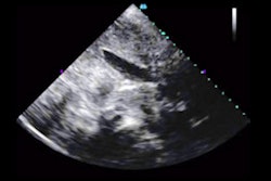
AI can walk ultrasound users through scans and enhance the image, ensuring the acquisition of high-quality and consistent scans that can be easily interpreted.
AI can also identify anatomy and anomalies on scans and make measurements to help readers interpret scans. AI can serve as a safeguard, ensuring that the radiologist does not miss any areas of concern on a scan, and as a second opinion, improving the radiologist's confidence in their diagnosis.
The chart below lists the importance of various AI tasks, as indicated in an Omdia survey of users.

According to the AIUS report, AI for ultrasound is versatile, with 60% of respondents' practices using AI in multiple clinical applications. AI utilization in general imaging was the most frequently reported clinical application, but Omdia expects AI utilization in point-of-care (POC), cardiology, and nontraditional applications to increase during the next few years as image libraries grow, enabling the development of more specialized algorithms.
The development of AI at the edge of care and in POC settings will drive integrated and cloud-based AI deployment in addition to the use of AI with portable medical imaging equipment.
As the virus continues to ravage healthcare systems around the world, healthcare providers are employing AI as an important tool to combat COVID-19. AI is being used to quickly screen and triage COVID-19 patients and identify symptoms on scans. This can help healthcare practices to limit the spread of the virus and quickly treat more patients.
While many healthcare providers are utilizing preexisting AI, many AI vendors and equipment manufacturers have developed and continue to develop AI software to aid in the identification and diagnosis of pulmonary and cardiovascular symptoms associated with COVID-19. The benefit and growing understanding of AI for COVID-19 is made clear by the accelerated development of software, increased funding, and easing of regulations.
While in many cases the advantage of using new technology is anecdotal, the benefit of AI is supported by several metrics tracked by healthcare administrators. According to the AIUS report, 85% of respondents reported that AI saved their practice time, 69% reported that the use of AI saved their practice money, 82% cited improved diagnostics as a top driver for AI adoption, 74% of respondents reported that AI increased their practices' utilization of ultrasound, and 49% reported that AI reduced the amount of training required for a technician to operate an ultrasound.
These results are significant because they demonstrate that an overwhelming majority of respondents see measurable improvements following implementation of AI for ultrasound.
Healthcare providers that have employed AI clearly understand the value of AI, and this is reflected by usage rates. The AIUS report identifies that 73% of respondents' practices use an AI feature at least once a day, with 40% of respondents reporting hourly usage.
This type of buy-in from the medical community is crucial to the widespread implementation of healthcare AI. AI vendors should continue to partner with equipment manufacturers, healthcare providers, and medical schools to identify key AI applications and to facilitate adoption.
Adam Davidson is a market research analyst with Omdia. The company's Artificial Intelligence for Healthcare Ultrasound Report Analysis was published in October 2020.
The comments and observations expressed are those of the author and do not necessarily reflect the opinions of AuntMinnie.com.
© 2020 Omdia. All rights reserved. Unauthorized reproduction prohibited.