Echocardiography revealed a wide range of serious heart abnormalities in patients hospitalized for COVID-19. The findings come from the first part of one of the largest studies on COVID-19 heart complications to date and were published on October 26 in the Journal of the American College of Cardiology.
Not only did many patients with COVID-19 experience heart injuries, but patients with abnormalities on cardiac ultrasound scans were also much more likely to experience worse outcomes, including death, mechanical ventilation, or admission to the intensive care unit (ICU).
"This is one of the first studies to provide detailed echocardiographic and electrocardiographic data in hospitalized patients with COVID-19 and laboratory evidence of myocardial injury," stated lead author Dr. Gennaro Giustino, a cardiology fellow at Mount Sinai Hospital in New York City.
Prior research has documented evidence of heart injury in people with COVID-19, but the mechanisms behind the cause of cardiac damage is not well understood. This is partially because of the intrinsic challenges of conducting safe, comprehensive scans during a pandemic.
The new study sought to shed light on heart injury in patients with COVID-19 through pathology, electrocardiogram (ECG), and data from transthoracic echocardiographic (TTE) exams. It included 305 patients hospitalized with the novel coronavirus at seven hospitals in New York City and Italy.
About two-thirds of the patients had biomarker evidence of myocardial injury, defined as elevated cardiac troponin levels. In statistical analysis, almost all echocardiographic characteristics were significantly associated with high serum troponin.
The TTE exams identified a wide range of cardiac abnormalities, including some linked to heart attack and heart failure. Right ventricular dysfunction was the most common abnormality experienced among patients with myocardial injury and COVID-19, followed by left ventricular dysfunction and left ventricular global dysfunction.
Patients with TTE and high troponin levels also had worse outcomes than those with normal echocardiographic findings. During the study period, 31% of patients with myocardial injury and echocardiographic abnormalities died, compared with 21% of patients with myocardial injuries but no echographic abnormalities and 5% of patients with normal troponin levels.
Furthermore, mortality levels remained increased for patients with myocardial injury and echocardiographic abnormalities, even after adjusting for all other major COVID-19 complications. The authors concluded that TTE findings in patients with troponin-positive COVID-19 syndromes provide useful prognostic information.
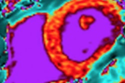
 Types of echocardiographic abnormalities in patients with COVID-19 and myocardial injury. Image courtesy of Mount Sinai Health System.
The study confirmed that COVID-19 can result in broad cardiovascular damage, although it's still unknown whether this occurs directly or indirectly as a result of the infection. Mechanisms behind heart injury could include cytokine-mediated myocardial damage, oxygen supply-demand imbalance, vascular thrombosis, and direct viral invasion.
It's important to note that the authors didn't use a systematic basis to select which patients to perform TTE, so only patients at increased cardiac risk may have undergone echocardiograms. It's also unknown what the lasting effects of these heart abnormalities may be, noted corresponding author Dr. Martin Goldman, from the Icahn School of Medicine at Mount Sinai.
"Because this is a new disease, we plan on following these patients closely using imaging to evaluate the evolution and hopefully resolution of these cardiac issues," stated Goldman.