The COVID-19 pandemic is driving an increase in the use of lung ultrasonography among physicians in Italy, according to the results of a small survey published in the Journal of Ultrasound in Medicine.
More physicians said they're using ultrasound equipment now than before the pandemic, and experienced physicians are performing more lung ultrasound scans than ever before. The findings support anecdotal evidence that COVID-19 has increased interest in ultrasound, including among clinicians with no prior ultrasound experience.
"Thanks to online resources, many operators could e-learn and apply the technique," wrote the authors, Dr. Allessandro Zanforlin and Dr. Francesco Tursi, who are both members of the Italian thoracic ultrasound academy behind the survey (July 16, 2020, Journal of Ultrasound in Medicine).
The informal, one-week-long survey was conducted by Italian ultrasound society Academia di Ecografia Thoracia after the group noticed its membership spiked from 1,700 members in February to 4,000 members in May. The academy promoted its survey on social media and received 123 responses.
Survey respondents worked in a variety of settings, including on COVID-19 floors (34%), in intensive care units (31%), and in the emergency department (20%). The majority said they used lung ultrasound to monitor pneumonia (63%) and screen for COVID-19 (60%).
A total of 14% of respondents started using lung ultrasound exclusively because of the COVID-19 pandemic. Among these respondents, 81% said they learned how to perform lung ultrasound scans by following video tutorials or participating in webinars. The remaining 19% of respondents gained experience through expert mentoring or local courses.
As the number of patients with COVID-19 surged at hospitals in Italy, so too did the number of lung ultrasound exams. Respondents said the number of daily chest scans increased from an average of three per day before the pandemic to seven per day during the outbreak. For so-called "expert operators" with at least five years of lung ultrasound experience, the number of exams rose from five per day to nine per day.
The majority of respondents also said their lung ultrasound exams had increased in quality (58%) and accuracy (66%) during the pandemic. These percentages were even higher for participants who took an online course or webinar, with 83% of these respondents saying their exams increased in both quality and accuracy.
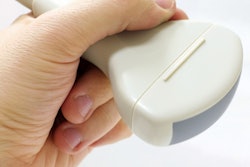
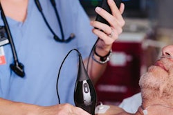
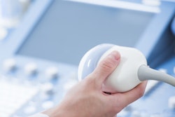
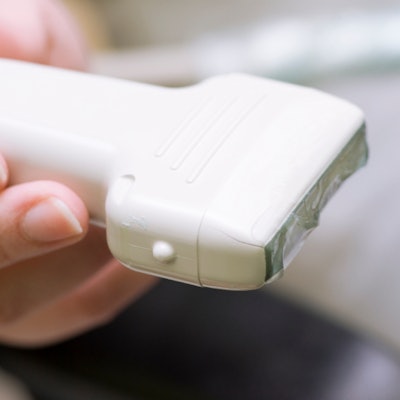
One factor driving the increase in lung ultrasound exams could be the availability of additional equipment thanks to donations and emergency purchase approvals. More than half of respondents said they acquired new ultrasound equipment during the pandemic, namely portable wheeled systems (37%) or handheld/wireless systems (19%).
The survey adds to the evidence that ultrasound is becoming an invaluable tool for care teams, especially those in Italy, who are treating patients with the novel coronavirus. However, the increase in skilled operators and new equipment may mean the modality will remain prominent for lung imaging even after the pandemic is over.
"What we are learning from this pandemic is the importance of [lung ultrasound] in the diagnosis, evaluations, and monitoring of pneumonia, which, in the hands of many physicians ... could improve the quality of the treatment of respiratory patients," the authors concluded.