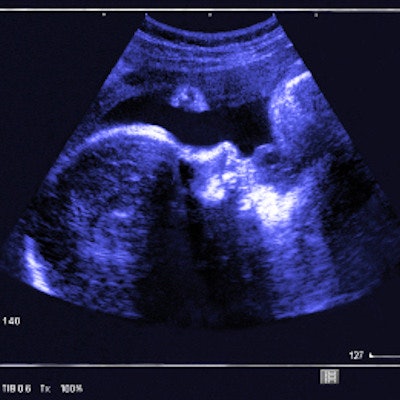
Researchers from Germany have received funding from the country's ministry of health to create an app that guides at-home ultrasound scans for pregnant patients. The app is part of a 2.5-year project to digitize maternal health in the country.
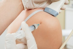
The project provides funding of 3.2 million euros ($3.5 million U.S.) to pay for a 500-person clinical trial with ob/gyn patients. The women will receive portable ultrasound probes, contraction monitors, and smartwatches or other smart devices to monitor their pregnancies. The researchers will develop a corresponding smartphone app to record, analyze, and send data to specialists.
Ideally, the app and accompanying tools will help address the doctor shortage in Germany. The country had more than 1,000 maternity departments in 1991, the researchers noted in a press release. Today there are only about 700.
"Our goal is not to replace personal contact to doctors and midwives and abandon expectant mothers to technology," stated Dr. Patrick Stelzl, deputy senior consultant for the project's medical aspects. "If we can reduce the time and effort required for routine examinations, we will then have more resources to deal intensively with complicated pregnancies."








![Examples of ultrasound findings and techniques. (A) Images in a 39-year-old male patient with a mass in the left thigh. The mass is heterogeneous on the B-mode US image (compared with the patient in D) and showed increased microvascularity (superb microvascular imaging [SMI]) and shear-wave elastography (SWE) values. Undifferentiated pleomorphic sarcoma was diagnosed at biopsy (with pleomorphic rhabdomyosarcoma in surgical specimen). (B) Images in an 18-year-old male patient with a mass in the left leg. The mass is hypoechoic on the B-mode image, with no other findings suggestive of malignancy. The lesion is in contact with the cortex of the tibia, which is slightly irregular. CT revealed a doubtful anteromedial tibial erosion. The microvascular study demonstrated high vascularization, suggestive of malignancy. Periosteal Ewing sarcoma was diagnosed with both histologic and immunohistochemical confirmation. (C) Images in a 69-year-old female patient with a lump growing on the outside of the left leg. Multiple SWE examinations were performed (please note the high values obtained in the measurements, whereas the color map highlights the stiffness relative to adjacent tissues). SMI showed areas of increased vascularization to target for sampling. Undifferentiated spindle cell sarcoma was diagnosed at biopsy, with residual leiomyosarcoma in the surgical specimen after neoadjuvant therapy. (D) Images in a 56-year-old female patient with a mass in the right thigh. The mass is heterogeneous at both B-mode ultrasound (similar to patient A) and MRI (coronal T2-weighted spectral attenuated inversion recovery [SPAIR]; T1-weighted pre-contrast and postcontrast imaging), which even shows uptake after the administration of paramagnetic contrast material, which is traditionally suggestive of malignancy. Low values at SMI and elastography are suggestive of benignity. Spindle cell lipoma was diagnosed at biopsy, with atypical spindle cell lipomatous tumor in the surgical specimen.](https://img.auntminnie.com/mindful/smg/workspaces/default/uploads/2026/08/images-radiol250278fig2.APCFLSvX6p.jpg?auto=format%2Ccompress&fit=crop&h=112&q=70&w=112)


