A new ultrasound technique could help clinicians assess bone health and distinguish between tumors and healthy tissue, according to one study published in the Journal of the Acoustical Society of America and another in Physics in Medicine & Biology.
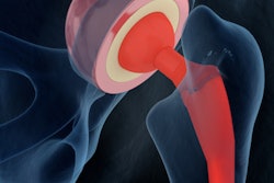
In one of the studies, researchers from North Carolina State University in Raleigh used a computational model to examine the rate at which ultrasound waves diffused from a bone site to assess pore density and the size of those pores (J Acoust Soc Am, August 8, 2019).
"One thing that's exciting about these techniques is that we've taken one of ultrasound's shortcomings and turned it to our advantage," said corresponding author Marie Muller, PhD, in a statement released by the university.
"In bone, for instance, sound waves will travel through the solid parts of the bone and then scatter whenever they hit a pore," she explained. "We found that this is a useful way to assess a bone's microstructure."
Although the technique is years away from clinical application, it could offer a way to monitor the bone health of patients, Muller said. She and her colleagues are currently conducting an in vivo study with human patients and an in vitro study with human bone.
"People can track their potential risk for osteoporosis without having to worry about the radiation exposure associated with x-rays," she said. "In addition, the technique could help researchers and healthcare providers determine the effect of osteoporosis treatment efforts."
Using ultrasound in this way to examine bone health could also be cost-effective, according to the university. The cost appears comparable to that of current ultrasound applications and less than the expense of high-resolution CT, though it's challenging to estimate due to the early nature of the research.
In the second study, Muller and colleagues used a similar ultrasound technique to distinguish between healthy soft tissue and solid tumors in rats. The rats' veins were injected with microbubbles, and the investigators then used ultrasound to track changes in the density of the microbubbles as they traveled through the animals' blood vessels (Phys Med Biol, May 31, 2019).
Compared with healthy tissue, tumors create denser, more complex networks of blood vessels, according to the researchers. Muller and colleagues found they could identify tumors by looking for areas where the microbubbles were slow to diffuse.
"We're excited about this finding, and are hoping to secure funding that would allow us to move forward with this line of research," she said.