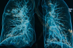
Lung ultrasound with a surface wave elastography (SWE) protocol is a viable tool for noninvasive evaluation of lung disease, giving clinicians another way to diagnose the condition besides CT or pulmonary function tests, according to a study published online March 5 in Radiology.
"Lung ultrasound has been increasingly used in the past decade for assessing lung disease," wrote a team led by Boran Zhou, PhD, of the Mayo Clinic in Rochester, MN. "Its advantages over other imaging modalities are speed, ease of use, portability, absence of ionizing radiation, and low cost."
Lung disease is evaluated by the amount of fibrosis in the organ, and pulmonary function tests have long been used in this assessment, Zhou's group noted. But these tests can have a wide range of normal values and are relatively insensitive for identifying early-stage disease. CT can also be used, but it requires radiation and can be expensive, the team wrote.
Zhou's group, therefore, sought to evaluate whether ultrasound could be an effective alternative to CT for quantifying lung surface stiffness or fibrosis by assessing the rate of surface wave propagation. The study included 91 participants with lung disease and 30 healthy subjects. Using pulmonary function tests, CT, and clinical evaluation, the researchers graded disease severity on a four-point scale:
- 0: No disease
- 1: Mild disease
- 2: Moderate disease
- 3: Severe disease
All study participants underwent lung ultrasound SWE. The researchers calculated optimal cutoff wave speed values for disease grades 0 through 3 using receiver operating characteristic (ROC) curve analysis.
Zhou's team found that lung ultrasound SWE had good sensitivity and specificity rates for distinguishing between healthy subjects and patients with any lung disease, and healthy subjects and those with mild disease. Since the highest ROC area under the curve values were found at 200 Hz, the investigators used a cutoff mean value for lung surface wave speed of 5.47 m/sec at the left lateral intercostal area at 200 Hz as the peak performance measure.
| Lung SWE for differentiating grades of lung disease | |||
| Lung SWE | Differentiating between no disease and grades 1-3 | Differentiating between grades 0-2 and grade 3 | Differentiating between no disease and grade 1 |
| Sensitivity | 92% | 50% | 88% |
| Specificity | 89% | 81% | 97% |
The results suggest that lung ultrasound with SWE protocol could be a useful screening tool for assessing lung disease in at-risk patients, and perhaps could even be used for long-term monitoring, according to Zhou and colleagues.
"The current clinical diagnostic standard technique for lung assessment is high-resolution CT, but it is costly, requires exposure to ionizing radiation, and typically is not performed serially," the group concluded. "In contrast, lung ultrasound SWE can be used to safely evaluate longitudinal changes in lung stiffness."