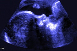
Abnormal results on prenatal ultrasound scans in women infected with the Zika virus may help predict how the virus will affect the fetus after birth -- but they can't be considered definitive, according to a study published online December 28 in JAMA Network Open.
Yes, ultrasound can identify the potential for adverse outcomes in fetuses exposed to the virus, but it's not the be-all and end-all solution because many babies with normal prenatal ultrasound findings are born with complications from Zika, wrote a team led by Dr. Jose Paulo Pereira Jr. of the Instituto Nacional de Saúde da Mulher, da Criança e do Adolescente in Rio de Janeiro.
"More than half of the patients [in our study] with abnormal results on neonatal examinations had no structural findings on prenatal ultrasonography. ... Our results suggest that prenatal ultrasonography may have a limited ability to provide reassurance of a normal neonatal outcome in maternal Zika virus infection," the authors wrote.
Literature on whether prenatal ultrasound can help determine how babies will be affected by the Zika virus has mostly included cohorts of babies identified pre- or postnatally with microcephaly. But Zika can cause other complications in the central nervous system, such as calcifications, ventriculomegaly, cerebellar vermis hypoplasia, and agenesis of the corpus callosum, according to the investigators.
Pereira's team sought to investigate the association between prenatal ultrasound findings and newborn outcomes among pregnant women infected with the virus. The study included 92 mother-baby pairs; the median gestational age of the babies at delivery was 38.6 weeks. Abnormal findings included a range of central nervous system irregularities, as well as other conditions such as fetal growth restriction, arthrogryposis, fluid abnormalities, placentomegaly, and macrosomia.
Of the 92 mother-baby pairs, 55 (60%) had normal results on prenatal ultrasound and 37 (40%) had abnormal results. Of the 55 pairs with normal results, 23 newborns had an adverse outcome (42%); of the 37 pairs with abnormal results, one fetus died at 36 weeks, and 21 of the remaining 36 had an abnormal outcome (58%).
"We found that major Zika virus-associated abnormalities observed on prenatal ultrasonography were associated with a sixfold to 27-fold increase in the odds of composite adverse neonatal outcomes of abnormal results on neonatal examination or postnatal neuroimaging," the authors wrote. They defined composite adverse neonatal outcomes as perinatal death, an abnormal finding on newborn exam, or an abnormal finding on postbirth neuroimaging.
| Association between ultrasound findings and adverse birth outcomes | |||
| Ultrasound result | Neonatal exam | Postnatal neuroimaging | Composite neonatal outcome |
| Zika virus-associated abnormal finding | |||
| Normal result | 3.9% | 4.4% | 2.1% |
| Abnormal result | 22% | 30.4% | 22.2% |
| Adjusted odds ratio | 11.6 | 6.7 | 27.2 |
| Central nervous system abnormality | |||
| Normal result | 3.9% | 2.2% | 2.1% |
| Abnormal result | 19.5% | 30.4% | 20% |
| Adjusted odds ratio | 11 | 13.9 | 27.2 |
An abnormal result of any type on prenatal ultrasound had a sensitivity of 48.9% and a specificity of 68.1%. For a major abnormal result that was specifically associated with Zika, sensitivity was 22.2%, specificity was 97.9%, positive predictive value was 90.9%, and negative predictive value was 56.8%.
The study shows that more research is necessary, according to Pereira and colleagues.
"In the setting of confirmed Zika virus infection in pregnancy, prenatal ultrasonography is a useful tool for anticipating an association with adverse neonatal outcomes, but its negative predictive value ... is low," they concluded.