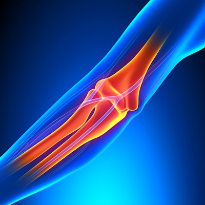
Real-time sonoelastography is a useful tool for diagnosing medial epicondylitis, or golfer's elbow, according to research presented by a team from South Korea at the recent RSNA 2017 meeting in Chicago.
Although the condition tends to be identified through a physical exam, its diagnosis is often confirmed by ultrasound imaging, especially in cases that remain unclear, said presenter Dr. Seok Hahn of Haeundae Hospital at Inje University in Busan. The fact that ultrasound is a viable tool for this indication is good news for both patients and physicians, as the modality is reliable, noninvasive, and inexpensive, Hahn told session attendees.
"Ultrasound is widely used to confirm the diagnosis and to evaluate the extent of medial epicondylitis because it is reliable, readily available, and inexpensive," he said. "Real-time sonoelastography has been applied to a wide range of musculoskeletal imaging applications, and it provides immediate and comprehensive information about tissue elasticity through color-coded mapping images."
Repetitive stress
Medial epicondylitis is caused by repetitive valgus stress, or overuse of flexor and pronator muscles that originate on the medial aspect of the elbow.
"In its early stages, medial epicondylitis presents as pain and functional impairment of the upper extremity," Hahn said. "If not diagnosed early, it can cause persistent pain and weakness at the elbow and require surgery."
To explore the technology's effectiveness in asymptomatic patients as well as those with suspected medial epicondylitis, Hahn and colleagues conducted a study that included grayscale sonography and compression-based real-time color sonoelastography imaging from 47 elbows in 43 patients performed between July 2016 and March 2017. Patients were divided into two groups: those with suspected medial epicondylitis and those with no symptoms.
On grayscale sonography, the researchers evaluated the presence of swelling, cortical irregularity, hypoechogenicity, tears, and calcification in the common flexor tendon. After this exam, the group performed real-time sonoelastography, focusing on the same field-of-view, and with manual compression. Hahn and colleagues then calculated the strain ratio based on the average strain measured in two regions of interest: the lesion in the common flexor tendon and, for reference, an adjacent normal tendon of similar depth.
Finally, to evaluate the diagnostic performance of real-time sonoelastography between the two patient groups, the researchers calculated receiving operating characteristic (ROC) curves for the different features on ultrasound that might predict golfer's elbow and compared them with the clinical diagnosis of medial epicondylitis as the reference standard.
Of the 43 patients, 17 (20 elbows) were diagnosed with medial epicondylitis. The strain ratio on elastography was the best predictor for medial epicondylitis, with an ROC of 0.97, while hypoechogenicity followed at 0.833, according to the researchers. On the other hand, clinical features including swelling, calcification, and tears showed no statistically significant differences between the two patient groups.
| Ultrasound features on elastography that predict golfer's elbow | |
| Ultrasound feature | ROC |
| Strain ratio | 0.968 |
| Hypoechogenicity | 0.833 |
| Elastographic grade | 0.814 |
| Cortical irregularity | 0.694 |
| Calcification | 0.631 |
"Elastographic grade showed good performance and strain ratio showed excellent performance for the diagnosis of medial epicondylitis," Hahn said.
Helpful tool
Although the study had some limitations, including the fact that the group set the clinical diagnosis of medial epicondylitis as the diagnostic standard, rather than pathology results, its findings show promise, according to Hahn.
"Real-time color sonoelastography that can obtain both elastographic grade and strain ratio makes it a valuable supplementary diagnostic tool in the diagnosis of this condition," he concluded.



![Examples of ultrasound findings and techniques. (A) Images in a 39-year-old male patient with a mass in the left thigh. The mass is heterogeneous on the B-mode US image (compared with the patient in D) and showed increased microvascularity (superb microvascular imaging [SMI]) and shear-wave elastography (SWE) values. Undifferentiated pleomorphic sarcoma was diagnosed at biopsy (with pleomorphic rhabdomyosarcoma in surgical specimen). (B) Images in an 18-year-old male patient with a mass in the left leg. The mass is hypoechoic on the B-mode image, with no other findings suggestive of malignancy. The lesion is in contact with the cortex of the tibia, which is slightly irregular. CT revealed a doubtful anteromedial tibial erosion. The microvascular study demonstrated high vascularization, suggestive of malignancy. Periosteal Ewing sarcoma was diagnosed with both histologic and immunohistochemical confirmation. (C) Images in a 69-year-old female patient with a lump growing on the outside of the left leg. Multiple SWE examinations were performed (please note the high values obtained in the measurements, whereas the color map highlights the stiffness relative to adjacent tissues). SMI showed areas of increased vascularization to target for sampling. Undifferentiated spindle cell sarcoma was diagnosed at biopsy, with residual leiomyosarcoma in the surgical specimen after neoadjuvant therapy. (D) Images in a 56-year-old female patient with a mass in the right thigh. The mass is heterogeneous at both B-mode ultrasound (similar to patient A) and MRI (coronal T2-weighted spectral attenuated inversion recovery [SPAIR]; T1-weighted pre-contrast and postcontrast imaging), which even shows uptake after the administration of paramagnetic contrast material, which is traditionally suggestive of malignancy. Low values at SMI and elastography are suggestive of benignity. Spindle cell lipoma was diagnosed at biopsy, with atypical spindle cell lipomatous tumor in the surgical specimen.](https://img.auntminnie.com/mindful/smg/workspaces/default/uploads/2026/08/images-radiol250278fig2.APCFLSvX6p.jpg?auto=format%2Ccompress&fit=crop&h=100&q=70&w=100)


![Examples of ultrasound findings and techniques. (A) Images in a 39-year-old male patient with a mass in the left thigh. The mass is heterogeneous on the B-mode US image (compared with the patient in D) and showed increased microvascularity (superb microvascular imaging [SMI]) and shear-wave elastography (SWE) values. Undifferentiated pleomorphic sarcoma was diagnosed at biopsy (with pleomorphic rhabdomyosarcoma in surgical specimen). (B) Images in an 18-year-old male patient with a mass in the left leg. The mass is hypoechoic on the B-mode image, with no other findings suggestive of malignancy. The lesion is in contact with the cortex of the tibia, which is slightly irregular. CT revealed a doubtful anteromedial tibial erosion. The microvascular study demonstrated high vascularization, suggestive of malignancy. Periosteal Ewing sarcoma was diagnosed with both histologic and immunohistochemical confirmation. (C) Images in a 69-year-old female patient with a lump growing on the outside of the left leg. Multiple SWE examinations were performed (please note the high values obtained in the measurements, whereas the color map highlights the stiffness relative to adjacent tissues). SMI showed areas of increased vascularization to target for sampling. Undifferentiated spindle cell sarcoma was diagnosed at biopsy, with residual leiomyosarcoma in the surgical specimen after neoadjuvant therapy. (D) Images in a 56-year-old female patient with a mass in the right thigh. The mass is heterogeneous at both B-mode ultrasound (similar to patient A) and MRI (coronal T2-weighted spectral attenuated inversion recovery [SPAIR]; T1-weighted pre-contrast and postcontrast imaging), which even shows uptake after the administration of paramagnetic contrast material, which is traditionally suggestive of malignancy. Low values at SMI and elastography are suggestive of benignity. Spindle cell lipoma was diagnosed at biopsy, with atypical spindle cell lipomatous tumor in the surgical specimen.](https://img.auntminnie.com/mindful/smg/workspaces/default/uploads/2026/08/images-radiol250278fig2.APCFLSvX6p.jpg?auto=format%2Ccompress&fit=crop&h=112&q=70&w=112)