Are you having a hard time classifying a renal lesion on CT, MRI, or conventional ultrasound? Contrast-enhanced ultrasound (CEUS) can be a powerful problem-solving tool for evaluating these indeterminate lesions, according to research published in the September issue of the Journal of Ultrasound in Medicine.
In a retrospective review, a team of researchers from the University of Alabama at Birmingham (UAB) found that CEUS could classify more than 95% of renal lesions deemed to be indeterminate on prior imaging studies. What's more, the method yielded 100% sensitivity and more than 85% specificity (J Ultrasound Med, September 2017, Vol. 36:9, pp. 1819-1827).
"Contrast-enhanced US is useful in differentiating benign from neoplastic lesions and can thus guide patient treatment," wrote the researchers led by Dr. Jessica Zarzour.
Indeterminate lesions
Small focal renal lesions less than 4 cm in size are often incidentally detected on CT, MRI, or conventional ultrasound. All three modalities can have challenges in characterizing these lesions, however. Although capable of distinguishing between cystic and solid lesions, conventional unenhanced ultrasound is unable to detect lesion enhancement -- an essential capability needed to determine the risk of malignancy in complex cysts, according to the authors.
Lesions discovered incidentally on a single-phase or unenhanced CT or MRI study may also be indeterminate; many small renal tumors show avid enhancement while other hypovascular tumors such as papillary renal cell carcinomas only demonstrate mild enhancement. Even on contrast-enhanced CT, though, pseudoenhancement can lead to false-positive findings in small renal lesions. Furthermore, false-negative results can occur in papillary renal cell carcinomas, which may show minimal enhancement.
On contrast-enhanced MRI, the detection of small enhancing components within a cystic lesion can also be challenging and possibly indeterminate, according to the authors. As an added complicating factor in evaluating these cases, patients with decreased renal function aren't able to receive contrast-enhanced CT or MRI due to the risk of renal damage or nephrogenic systemic fibrosis.
Consequently, contrast ultrasound is increasingly being utilized to evaluate renal lesions. Ultrasound contrast agents are based on microbubbles, which are not nephrotoxic and have a low incidence of side effects.
"A strength of contrast-enhanced US is its ability to be used in patients with impaired renal function or history of a contrast agent allergy," the authors wrote.
The utility of CEUS
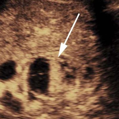
 CEUS showed a mixed solid and cystic lesion with enhancement of the solid component (arrow). This lesion was classified as potentially malignant, class B, and was a clear cell renal cell carcinoma on histopathologic examination. Image courtesy of the Journal of Ultrasound in Medicine.
The researchers sought to assess the utility of contrast ultrasound for characterizing lesions that were previously indeterminate on CT, MRI, or conventional ultrasound. They retrospectively evaluated 116 consecutive patients with 134 renal lesions who were evaluated with CEUS at their tertiary care hospital from 2006 to 2015. Of the 134 lesions, 106 were indeterminate on prior imaging, which included unenhanced CT, contrast-enhanced CT, unenhanced MRI, contrast-enhanced MRI, or unenhanced conventional ultrasound.
After first receiving conventional grayscale ultrasound to localize the renal lesion and determine the best plane for the contrast study, the CEUS exams were performed using a low mechanical index on either an iU22 (Philips Healthcare) or Epiq 7 (Philips) ultrasound scanner equipped with a C5-2 or C9-2 transducer. Once they had completed a safety questionnaire to assess for any potential contraindication to receiving microbubble contrast agents, the patients received intravenous bolus injections of the Definity (Lantheus Medical Imaging) or SonoVue (Bracco Diagnostics) ultrasound contrast agents. Most patients received one to three doses of contrast material, depending on if there were multiple lesions or if additional imaging was necessary, according to the researchers.
All studies were performed and immediately interpreted by an abdominal imaging fellowship-trained radiologist with four to 20 years of experience in contrast ultrasound. A fellowship-trained radiologist with four years of CEUS experience then retrospectively reviewed the studies on an ultrasound PACS (Imorgon Medical).
High classification rate
The radiologists were able to classify 90 (95.7%) of the 94 previously indeterminate renal lesions successfully. The remaining four indeterminate lesions included one case in the setting of acute hemorrhage and three with technical limitations due to a large body habitus and a small lesion diameter.
The researchers then evaluated the diagnostic performance of CEUS in the 41 patients who had a histologic diagnosis or who had received follow-up for more than one year. On follow-up ultrasound, any lesion that grew more than 20% in maximum diameter was considered to be malignant.
Contrast ultrasound yielded a high level of diagnostic performance for correctly classifying indeterminate renal lesions.
| Performance of contrast ultrasound for indeterminate renal lesions | |
| Sensitivity | 20/20 (100%) |
| Specificity | 18/21 (85.7%) |
| Positive predictive value | 20/23 (87%) |
| Negative predictive value | 18/18 (100%) |
| Accuracy | 37/41 (90.2%) |
The researchers acknowledged a number of limitations to their study, including its retrospective nature and the inclusion of a relatively low number of malignant lesions. In addition, only a subset of the lesions had pathologic confirmation. They also noted that renal cell carcinomas may remain stable in size for several years and would benefit from further follow-up.
"However, the benefit of using contrast-enhanced US to assess for enhancement in a renal lesion that was unable to be previously characterized is of great clinical importance and a powerful problem-solving tool," the group wrote.