Sunday, November 27 | 10:55 a.m.-11:05 a.m. | SSA02-02 | Room N228
Mammography technologists' performance with diagnostic automated breast ultrasound (ABUS) is comparable to that of sonographers, according to research being presented on Sunday morning at RSNA 2016.The study led by Dr. Richard Barr, PhD, a professor of radiology at Northeast Ohio Medical University in Rootstown, included 90 patients undergoing diagnostic ultrasound due to a palpable mass, an abnormality on mammography, a follow-up study, or breast discharge.
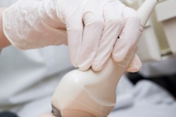
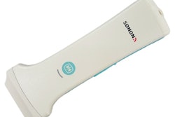
Barr's group compared the diagnostic accuracy of a handheld ultrasound exam and a single-volume ultrasound exam using an automated breast volume scanner positioned over the area of concern. The group also evaluated whether there was a performance difference between sonographers and mammography technologists.
All patients underwent both exams; they were randomized to have either handheld or automated ultrasound first. In addition, the automated ultrasound exams were randomized between a sonographer and a mammography technologist, and the person performing the second study was blinded to the results of the first exam.
Kappa agreement for finding pathology between automated breast and handheld ultrasound was 0.831, according to the researchers. The agreement between the two modalities was nearly the same when automated ultrasound was performed by a mammography technologist (0.858) as when it was performed by a sonographer (0.803) -- although this result was not statistically significant (p = 0.47).
There was no statistical difference between readers for the area under the curve (AUC) for lesion characterization for both automated and handheld ultrasound, leading Barr's group to conclude that handheld and automated breast ultrasound performed by a mammography technologist or a sonographer are statistically equivalent for diagnostic workup.




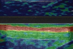


![Examples of ultrasound findings and techniques. (A) Images in a 39-year-old male patient with a mass in the left thigh. The mass is heterogeneous on the B-mode US image (compared with the patient in D) and showed increased microvascularity (superb microvascular imaging [SMI]) and shear-wave elastography (SWE) values. Undifferentiated pleomorphic sarcoma was diagnosed at biopsy (with pleomorphic rhabdomyosarcoma in surgical specimen). (B) Images in an 18-year-old male patient with a mass in the left leg. The mass is hypoechoic on the B-mode image, with no other findings suggestive of malignancy. The lesion is in contact with the cortex of the tibia, which is slightly irregular. CT revealed a doubtful anteromedial tibial erosion. The microvascular study demonstrated high vascularization, suggestive of malignancy. Periosteal Ewing sarcoma was diagnosed with both histologic and immunohistochemical confirmation. (C) Images in a 69-year-old female patient with a lump growing on the outside of the left leg. Multiple SWE examinations were performed (please note the high values obtained in the measurements, whereas the color map highlights the stiffness relative to adjacent tissues). SMI showed areas of increased vascularization to target for sampling. Undifferentiated spindle cell sarcoma was diagnosed at biopsy, with residual leiomyosarcoma in the surgical specimen after neoadjuvant therapy. (D) Images in a 56-year-old female patient with a mass in the right thigh. The mass is heterogeneous at both B-mode ultrasound (similar to patient A) and MRI (coronal T2-weighted spectral attenuated inversion recovery [SPAIR]; T1-weighted pre-contrast and postcontrast imaging), which even shows uptake after the administration of paramagnetic contrast material, which is traditionally suggestive of malignancy. Low values at SMI and elastography are suggestive of benignity. Spindle cell lipoma was diagnosed at biopsy, with atypical spindle cell lipomatous tumor in the surgical specimen.](https://img.auntminnie.com/mindful/smg/workspaces/default/uploads/2026/08/images-radiol250278fig2.APCFLSvX6p.jpg?auto=format%2Ccompress&fit=crop&h=112&q=70&w=112)


