A team from the University of California, Los Angeles (UCLA) has successfully used ultrasound to jump-start a patient's brain as he recovered from a coma, restoring full consciousness and language comprehension within three days of the procedure.
Before receiving a technique called low-intensity focused ultrasound pulsation, the patient had shown only minimal signs of consciousness and of understanding speech, according to a group led by Martin Monti, PhD, an associate professor of psychology and neurosurgery.
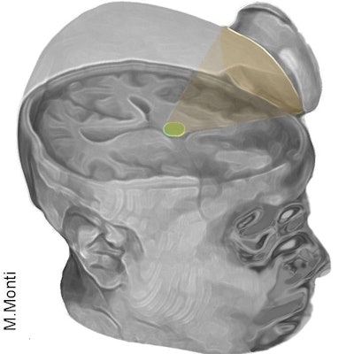
 The low-intensity focused ultrasound pulsation technique excites neurons in the thalamus. Image courtesy of Martin Monti, PhD.
The patient's responses improved measurably a day after the treatment, and he could reliably communicate by nodding his head or shaking his head within three days. What's more, the patient could even make a fist-bump gesture to say goodbye to one of his doctors, the UCLA researchers said. They described their experience in a study published online July 22 in Brain Stimulation.
The ultrasound device, which was developed by focused ultrasound technology developer BrainSonix, was used to excite neurons in the thalamus -- the brain's central hub for processing information. BrainSonix was founded by study co-author Dr. Alexander Bystritsky, who is also a professor of psychiatry and biobehavioral sciences at UCLA's Semel Institute for Neuroscience and Human Behavior.
The device, which is about the size of a coffee-cup saucer, creates a small sphere of acoustic energy that can be targeted at different brain regions to excite tissue, the researchers said. In their study, they placed it by the side of the man's head and activated it 10 times for 30 seconds each over a 10-minute period.
The researchers focused on the thalamus because thalamus performance is typically diminished in people whose mental function is deeply impaired after a coma, according to the group. Previously, the only way to excite neurons in the thalamus was via deep brain stimulation, a risky surgical procedure that involves implanting electrodes directly inside the thalamus, Monti said.
The UCLA group now plans to test the procedure on several more people this fall at the Ronald Reagan UCLA Medical Center. Testing will be performed in partnership with the UCLA Brain Injury Research Center and funded in part by the Dana Foundation and the Tiny Blue Dot Foundation.
If the technology helps others recovering from a coma, the researchers envision that it could eventually be used to build an inexpensive portable device and even possibly be incorporated into a helmet. It might even offer -- for the first time -- a treatment for patients in a vegetative or minimally conscious state, Monti said in a statement.