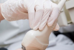
NEW YORK CITY - Breast ultrasound screening in women with dense breasts can identify invasive cancers not seen on mammography, and performance improves with experience, according to a presentation on Sunday at the American Institute of Ultrasound in Medicine (AIUM) annual meeting.
Now in its second year of providing breast ultrasound screening after adoption of New York's breast density notification law, Elizabeth Wende Breast Care in Rochester, NY, has been able to detect more cancers and perform fewer biopsies based on ultrasound results, for example.
"In the evaluation of screening breast ultrasound in women with dense breast tissue, we have found an increase in [positive predictive value] as well as an increase in our cancer detection rate, with a reduction in our biopsy rate," said presenter Dr. Stamatia Destounis.
She discussed the outpatient breast center's experience in providing screening breast ultrasound services for women with dense breasts during a scientific session at AIUM 2016.
Improved cancer detection
The researchers wanted to determine the effect of screening breast ultrasound on breast cancer detection in women with mammographically dense breasts, so they retrospectively reviewed screening ultrasound examinations performed at their institution between June 1, 2014, and August 31, 2015. New York's breast notification law took effect in 2013, mandating that all women be notified of their breast density in the summary report sent with their mammogram results and recommendations.
 Dr. Stamatia Destounis of Elizabeth Wende Breast Care.
The researchers calculated the total number of screening mammograms, screening ultrasound scans performed in dense breasts, procedures performed, and biopsy results including demographic data on those patients diagnosed with cancer.
Of the 93,817 screening mammograms performed, 36,341 (39%) were designated as having heterogeneously dense or extremely dense breast tissue. There were 4,900 screening ultrasound exams performed on 4,772 patients.
Among the patients who received screening ultrasound exams, 51 (1%) later received biopsies and other procedures. There were 12 cancers and one atypical lesion, yielding a positive predictive value of 24%.
The detected cancers were all invasive, with an average lesion size of 1.4 cm at excision. Eleven were node-negative, Destounis said. Ultrasound had a 99% specificity rate for the 4,900 screening ultrasounds that were performed.
"The study results ... demonstrate that screening breast ultrasound in women with dense breast tissue can detect otherwise occult malignancy, with a low recall rate and a reasonable [positive predictive value] for biopsy," she said.
Experience helps
After comparing the performance of the second year at Elizabeth Wende with the first year of providing screening ultrasounds, a number of improvements were evident.
| Screening ultrasound performance in women with dense breasts | ||
| January 1, 2013, to May 31, 2014 | June 1, 2014, to August 31, 2015 | |
| Screening mammograms | 102,841 | 93,817 |
| No. of women designated with dense tissue | 47,017 | 36,341 |
| No. of screening ultrasounds | 971 | 4,900 |
| Biopsy rate from screening ultrasound | 2.6% | 1% |
| Cancer detection rate from screening ultrasound | 2.1 per 1,000 | 2.5 per 1,000 |
| Positive predictive value of screening ultrasound | 8% | 24% |
"We got better at doing it the following year," Destounis said.
The increase in positive predictive value reflected the learning curve for breast ultrasound screening, as the institution performed fewer biopsies in the second year, according to Destounis.
"We got better at what we were targeting for biopsy," she said. "So we did less false-positive biopsies and our [positive predictive value] increased."
The number of women designated with dense breast tissue also declined in the second year of the program.
She also noted that while the radiologists had initially interpreted breast density themselves as they read the mammograms, the practice's adoption of breast density classification software led to a decrease in the number of women designated as having dense breasts.
"We found out that we overcalled [breast density] a little, so the percentage of actual dense breasts in our facility actually went down a little bit as we went to the automated system," she said.