The American Institute of Ultrasound in Medicine (AIUM) has released guidelines for improving the use of ultrasound in obstetrics and gynecology, in both clinical practice and residency training.
Published simultaneously on January 3 in the American Journal of Obstetrics and Gynecology, the Journal of Ultrasound in Medicine, and Ultrasound in Obstetrics and Gynecology, the guidance is the result of a forum AIUM held in Boston in October 2016.
The forum, Beyond Ultrasound First, was led by Dr. Beryl Benacerraf from Brigham and Women's Hospital and Harvard Medical School. It included representatives from professional associations; the imaging, ob/gyn, and emergency medicine communities; and government agencies, insurers, and industry.
Although most obstetricians and gynecologists agree that ultrasound should be the first-choice imaging method for their patients, there is inconsistency in its use and the level of competency with which it is performed, according to the AIUM. The forum explored topics such as understanding the role of ultrasound in clinical imaging, minimizing false-positive and false-negative findings, standardizing training, and understanding the perspectives of payors toward the modality.
"Improving the quality of ultrasound examinations will have a substantial impact on patient care and healthcare costs," Benacerraf said in an AIUM statement. "To our knowledge, this is the first time that a multisociety curriculum and competency assessment tools have been created for residency training in clinical imaging."
Better curriculum
Dr. Alfred Abuhamad of Eastern Virginia Medical School in Norfolk led the AIUM task force charged with developing an ob/gyn ultrasound curriculum for residents. The group's report outlines essential topics in medical ultrasound training, a skill development framework, and a competency assessment that involves the evaluation of still ultrasound images, movie clips, and real-time scanning.
"Ensuring optimal training of ultrasound in residency programs will result in improved quality of ultrasound examinations in clinical practice," Abuhamad said.
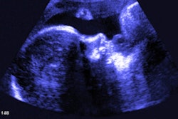
 Biparietal diameter along with the list of criteria for competency: A teaching example from the curriculum that describes how an important measurement of a developing fetus is made. Image courtesy of the American Journal of Obstetrics and Gynecology.
Ultrasound is much more operator-dependent than other imaging modalities, and obstetric ultrasound in particular can be challenging, due to the small size of fetal organs, according to the AIUM. This makes training and competency assessment all the more important, Benacerraf said.
"Ultrasound can be immensely informative, but it requires more skill and training than just pushing a button," she said. "Those that make the effort to master the modality will reap rich rewards for their patients, trainees, and themselves."