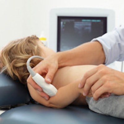
Do patients really want to get imaging findings directly from radiologists? When it comes to the parents of children receiving ultrasound studies, they almost always do, according to a study published in the October issue of the American Journal of Roentgenology.
Researchers from Stanford University School of Medicine implemented radiologist-patient consultations after routine outpatient pediatric ultrasound studies at Lucile Packard Children's Hospital. They found that more than 90% of parents indicated they would prefer to meet directly with the radiologist at every visit going forward. The parents also reported significant improvements in parental anxiety and satisfaction. What's more, they had a much better understanding afterward of the radiologist's role.
"On the basis of our results, we strongly advocate that consultation sessions for outpatient pediatric ultrasound be offered routinely to parents," wrote Dr. Evan Zucker and colleagues.
Radiologist-patient interactions
Radiologist-patient consultations have been proposed as a way to boost the visibility of the specialty, and they have been shown in a number of studies in the literature to be preferred by patients. While pediatric radiology is considered to be a more patient-oriented subset of radiology than others, the researchers said they were not aware of any previous studies that assessed radiologist-patient or radiologist-parent interactions for pediatric imaging.
As a result, they set out to implement such consultation sessions for routine pediatric ultrasound -- with immediate disclosure of results, if desired -- to determine whether such interactions increase visit satisfaction, decrease anxiety, and grow understanding of the radiologist's role (AJR, October 2015, Vol. 205:4, pp. 822-826).
Parents who were chaperoning any outpatient pediatric ultrasound were eligible for the study. Participants completed surveys before and after the ultrasound exams, and they met with a pediatric radiologist before the second survey. They could opt out and then request or decline the consultation, according to the researchers.
Of the 77 subjects (55 women, 22 men; mean age, 36.8 ± 8.7 years; age range, 18-62 years) who participated, 71 (92%) chose to speak with a radiologist when given the opportunity. One did not answer the first survey's questions on age, race, or ethnicity.
Among those who received radiologist consultations, the subjects rated overall visit experience as follows:
- Excellent: 56 (80%)
- Good: 13 (19%)
- Fair: 1 (1%)
The subjects also rated the value of the radiologist consultation:
- Very helpful: 55 (77%)
- Moderately helpful: 12 (17%)
- Mildly helpful: 3 (4%)
- Not helpful: 1 (1%)
They rated radiologist interaction as follows:
- Excellent: 50 (70%)
- Good: 20 (28%)
- Poor: 1 (1%)
Multivariate analysis did not find a statistically significant predictor of patient satisfaction, according to the authors.
Prior to consultation with the radiologist, the subjects had a mean anxiety score of 2.0 ± 1.0 on a scale of 1 (lowest) to 4 (highest). The score dropped to 1.5 ± 0.8 after consultation -- a statistically significant improvement (p < 0.001).
After the radiologist consultation, 60 (88%) of 68 respondents correctly described the radiologist's role, up from 46 (68%) prior to consultation. This increase was also statistically significant (p < 0.001).
Appreciative of the opportunity
In addition, 64 (91%) of 70 respondents indicated they would prefer to speak with a radiologist during every visit, while five (7%) said they would prefer to receive consultation during future visits only upon request and one (1%) did not want consultation in the future. The researchers also noted that write-in comments on the surveys were mostly positive.
"Parents appreciated the opportunity to speak with a radiologist, receive immediate results, and have their questions answered," the authors wrote. "Some described a decrease in anxiety. They further described staff, including the technologists, as excellent, friendly, knowledgeable, understanding, and compassionate."
The researchers noted that consultations for all outpatient ultrasound exams are currently provided at their clinical practice by radiologists when the patient or family requests it. A consultation is not offered for every examination due to limitations in staffing.
"In our experience, consultation does not excessively increase radiologist workload," the authors wrote. "Yet competing activities may have to wait until consultation sessions finish, causing some delay in traditional workflow processes. Nevertheless, we are reviewing options for reorganizing our ultrasound coverage so that we can offer this important and valued additional service in all cases."