Dear Ultrasound Insider,
In today's busy world, it's easy to get bogged down in the day-to-day grind and lose track of what's important. For sonographers, that can mean neglecting physical health and, consequently, experiencing wrist and forearm pain.
In the latest Making a Difference as a Sonographer column, echocardiographer Doug Wuebben shares his thoughts on the importance of setting priorities, and he also provides some tips and exercises from strength and conditioning coach Mark Roozen. This Insider Exclusive is available to you first as a subscriber.
In other features in your Ultrasound Community, research presented at the recent European League Against Rheumatism annual congress examined the benefits of ultrasound for fine-tuning the management of rheumatoid arthritis patients who are in remission. The modality was shown to identify subclinical disease activity in these patients, enabling them to benefit from more-intensive treatment. Click here for the details.
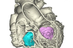
Michigan researchers have created a 3D printed heart that was generated using data from echocardiography and CT studies. International Editor Eric Barnes has our coverage, which you can access here.
Thanks to its ability to reliably distinguish between patients who have no or minimal fibrosis and those with severe fibrosis or cirrhosis, ultrasound elastography can obviate the need for liver biopsy in most cases, according to a multispecialty panel convened by the Society of Radiologists in Ultrasound. Learn more here.
Dr. Jason Birnholz also has returned with another column in his popular Practice of Ultrasound series. In the latest edition, Dr. Birnholz shares his exploration of ultraportable ultrasound technology.
Did you hear about the Australian woman who earlier this month pleaded guilty to providing fake keepsake ultrasound images to expectant mothers? Read all about it by clicking here.
Is there a topic you'd like to see covered? As always, please feel free to drop me a line.






![Examples of ultrasound findings and techniques. (A) Images in a 39-year-old male patient with a mass in the left thigh. The mass is heterogeneous on the B-mode US image (compared with the patient in D) and showed increased microvascularity (superb microvascular imaging [SMI]) and shear-wave elastography (SWE) values. Undifferentiated pleomorphic sarcoma was diagnosed at biopsy (with pleomorphic rhabdomyosarcoma in surgical specimen). (B) Images in an 18-year-old male patient with a mass in the left leg. The mass is hypoechoic on the B-mode image, with no other findings suggestive of malignancy. The lesion is in contact with the cortex of the tibia, which is slightly irregular. CT revealed a doubtful anteromedial tibial erosion. The microvascular study demonstrated high vascularization, suggestive of malignancy. Periosteal Ewing sarcoma was diagnosed with both histologic and immunohistochemical confirmation. (C) Images in a 69-year-old female patient with a lump growing on the outside of the left leg. Multiple SWE examinations were performed (please note the high values obtained in the measurements, whereas the color map highlights the stiffness relative to adjacent tissues). SMI showed areas of increased vascularization to target for sampling. Undifferentiated spindle cell sarcoma was diagnosed at biopsy, with residual leiomyosarcoma in the surgical specimen after neoadjuvant therapy. (D) Images in a 56-year-old female patient with a mass in the right thigh. The mass is heterogeneous at both B-mode ultrasound (similar to patient A) and MRI (coronal T2-weighted spectral attenuated inversion recovery [SPAIR]; T1-weighted pre-contrast and postcontrast imaging), which even shows uptake after the administration of paramagnetic contrast material, which is traditionally suggestive of malignancy. Low values at SMI and elastography are suggestive of benignity. Spindle cell lipoma was diagnosed at biopsy, with atypical spindle cell lipomatous tumor in the surgical specimen.](https://img.auntminnie.com/mindful/smg/workspaces/default/uploads/2026/08/images-radiol250278fig2.APCFLSvX6p.jpg?auto=format%2Ccompress&fit=crop&h=112&q=70&w=112)