Dear Ultrasound Insider,
Fetal ultrasound in the second or third trimester is thought to be pretty accurate for detecting situs inversus, a congenital condition in which the chest and abdominal organs are reversed. But all may not be as it seems.
In part 21 of his ongoing Practice of Ultrasound series, ultrasound luminary Dr. Jason Birnholz delves into situs inversus, explores the brain of the radiologist, and makes the case that ultrasound will benefit from a greater appreciation of molecular biology. The column is this issue's Insider Exclusive, which you have access to before our regular members.
In other featured articles in your Ultrasound Community, researchers from Boston Children's Hospital recently found that the combination of ultrasound and blood tests significantly outperformed ultrasound alone for detecting pediatric appendicitis. What's more, the duo could also lower the number of CT studies needed in these patients. Learn more by clicking here.
We also have a new article from echocardiographer Doug Wuebben and strength coach Mark Roozen in their popular Making a Difference as a Sonographer series. In their latest column, they show how foam rollers and other tools can be used in recovery exercises.
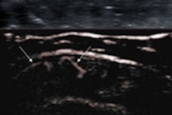
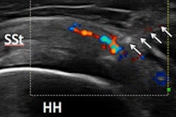
Meanwhile, researchers from the U.K. found that a new Doppler ultrasound technique could yield better visualization of low-grade inflammation in joints and tendons than power Doppler ultrasound. The added clinical information often influences patient management, according to the group.
An Italian team recently concluded that ultrasound-guided injections of platelet-rich plasma for rotator cuff tendinopathy could delay degenerative changes in the tendons and offer pain relief and functional improvement. Click here to learn more.
Finally, a study from the U.S. National Cancer Institute determined that prostate cancer biopsy performed with a targeted technique that fuses MR and ultrasound images was superior to traditional extended-sextant ultrasound-guided biopsy. Click here for all the findings.
Is there a topic you'd like to see covered? As always, please feel free to drop me a line.








![Examples of ultrasound findings and techniques. (A) Images in a 39-year-old male patient with a mass in the left thigh. The mass is heterogeneous on the B-mode US image (compared with the patient in D) and showed increased microvascularity (superb microvascular imaging [SMI]) and shear-wave elastography (SWE) values. Undifferentiated pleomorphic sarcoma was diagnosed at biopsy (with pleomorphic rhabdomyosarcoma in surgical specimen). (B) Images in an 18-year-old male patient with a mass in the left leg. The mass is hypoechoic on the B-mode image, with no other findings suggestive of malignancy. The lesion is in contact with the cortex of the tibia, which is slightly irregular. CT revealed a doubtful anteromedial tibial erosion. The microvascular study demonstrated high vascularization, suggestive of malignancy. Periosteal Ewing sarcoma was diagnosed with both histologic and immunohistochemical confirmation. (C) Images in a 69-year-old female patient with a lump growing on the outside of the left leg. Multiple SWE examinations were performed (please note the high values obtained in the measurements, whereas the color map highlights the stiffness relative to adjacent tissues). SMI showed areas of increased vascularization to target for sampling. Undifferentiated spindle cell sarcoma was diagnosed at biopsy, with residual leiomyosarcoma in the surgical specimen after neoadjuvant therapy. (D) Images in a 56-year-old female patient with a mass in the right thigh. The mass is heterogeneous at both B-mode ultrasound (similar to patient A) and MRI (coronal T2-weighted spectral attenuated inversion recovery [SPAIR]; T1-weighted pre-contrast and postcontrast imaging), which even shows uptake after the administration of paramagnetic contrast material, which is traditionally suggestive of malignancy. Low values at SMI and elastography are suggestive of benignity. Spindle cell lipoma was diagnosed at biopsy, with atypical spindle cell lipomatous tumor in the surgical specimen.](https://img.auntminnie.com/mindful/smg/workspaces/default/uploads/2026/08/images-radiol250278fig2.APCFLSvX6p.jpg?auto=format%2Ccompress&fit=crop&h=112&q=70&w=112)