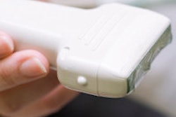
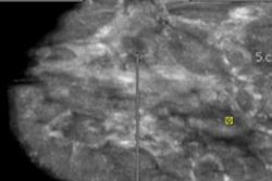
Radiologists show equal confidence in visualizing suspicious masses with automated breast ultrasound (ABUS) and handheld breast ultrasound, which could decrease the amount of handheld imaging used in diagnostic clinics, according to research presented at the recent RSNA 2013 meeting.
The benefits of ABUS include decreased interpretation time (some studies have found that ABUS takes only three minutes, while handheld US takes at least 20), which improves a department's throughput. But automated breast ultrasound also reduces operator variation, said Dr. Cherie Kuzmiak from the University of North Carolina at Chapel Hill.
"Mammography is the gold standard for breast cancer detection, but density is a major limitation to its sensitivity, while ultrasound is not limited by density," she told session attendees. "It's a sensitive modality, but traditionally it involves using a handheld transducer, which contributes to a significant amount of operator subjectivity and variation. We wanted to investigate how well radiologists visualize relevant features of lesions seen with automated breast volumetric scanning in comparison to handheld breast ultrasound in a population of women going to biopsy."
For the study, Kuzmiak and colleagues included 25 women scheduled to undergo a breast biopsy for at least one BI-RADS 4 or 5 lesion identified in a diagnostic setting. Each patient underwent imaging with a system that allowed both handheld breast ultrasound and automated breast volumetric scanning to be performed with the same imaging parameters.
The study included five experienced breast imaging radiologists, who compared the handheld and automated breast exams side by side. Each reader specified lesion type, size, and imaging features; BI-RADS score; and probability of malignancy for each lesion for each modality. They were then asked to compare the lesion characteristics of shape and margin between the two modalities using a seven-point confidence scale.
Of 30 biopsied lesions, all were masses. Seven (23%) were malignant and 23 (77%) were benign. Across all lesions regardless of size or final pathology, there was no significant difference between the two ultrasound modalities in the readers' BI-RADS classification, probability of malignancy, sensitivity, or specificity, the researchers found.
For malignant lesions, the reader confidence scores between the two modalities did not differ significantly; however, readers were more confident of their analysis of lesion shape and margins for nonmalignant cases with automated breast volumetric scanning.
"We attributed this to the fact that with automated breast ultrasound, the radiologist is provided with more information on multiple planes, similar to an MRI," Kuzmiak said.
Dedicated automated whole-breast ultrasound could reduce handheld breast imaging use, she concluded.
"Our other hope is that by decreasing the need for handheld ultrasound, the need for targeted ultrasound after screening exams could also decrease," she said.


![Examples of ultrasound findings and techniques. (A) Images in a 39-year-old male patient with a mass in the left thigh. The mass is heterogeneous on the B-mode US image (compared with the patient in D) and showed increased microvascularity (superb microvascular imaging [SMI]) and shear-wave elastography (SWE) values. Undifferentiated pleomorphic sarcoma was diagnosed at biopsy (with pleomorphic rhabdomyosarcoma in surgical specimen). (B) Images in an 18-year-old male patient with a mass in the left leg. The mass is hypoechoic on the B-mode image, with no other findings suggestive of malignancy. The lesion is in contact with the cortex of the tibia, which is slightly irregular. CT revealed a doubtful anteromedial tibial erosion. The microvascular study demonstrated high vascularization, suggestive of malignancy. Periosteal Ewing sarcoma was diagnosed with both histologic and immunohistochemical confirmation. (C) Images in a 69-year-old female patient with a lump growing on the outside of the left leg. Multiple SWE examinations were performed (please note the high values obtained in the measurements, whereas the color map highlights the stiffness relative to adjacent tissues). SMI showed areas of increased vascularization to target for sampling. Undifferentiated spindle cell sarcoma was diagnosed at biopsy, with residual leiomyosarcoma in the surgical specimen after neoadjuvant therapy. (D) Images in a 56-year-old female patient with a mass in the right thigh. The mass is heterogeneous at both B-mode ultrasound (similar to patient A) and MRI (coronal T2-weighted spectral attenuated inversion recovery [SPAIR]; T1-weighted pre-contrast and postcontrast imaging), which even shows uptake after the administration of paramagnetic contrast material, which is traditionally suggestive of malignancy. Low values at SMI and elastography are suggestive of benignity. Spindle cell lipoma was diagnosed at biopsy, with atypical spindle cell lipomatous tumor in the surgical specimen.](https://img.auntminnie.com/mindful/smg/workspaces/default/uploads/2026/08/images-radiol250278fig2.APCFLSvX6p.jpg?auto=format%2Ccompress&fit=crop&h=100&q=70&w=100)


![Examples of ultrasound findings and techniques. (A) Images in a 39-year-old male patient with a mass in the left thigh. The mass is heterogeneous on the B-mode US image (compared with the patient in D) and showed increased microvascularity (superb microvascular imaging [SMI]) and shear-wave elastography (SWE) values. Undifferentiated pleomorphic sarcoma was diagnosed at biopsy (with pleomorphic rhabdomyosarcoma in surgical specimen). (B) Images in an 18-year-old male patient with a mass in the left leg. The mass is hypoechoic on the B-mode image, with no other findings suggestive of malignancy. The lesion is in contact with the cortex of the tibia, which is slightly irregular. CT revealed a doubtful anteromedial tibial erosion. The microvascular study demonstrated high vascularization, suggestive of malignancy. Periosteal Ewing sarcoma was diagnosed with both histologic and immunohistochemical confirmation. (C) Images in a 69-year-old female patient with a lump growing on the outside of the left leg. Multiple SWE examinations were performed (please note the high values obtained in the measurements, whereas the color map highlights the stiffness relative to adjacent tissues). SMI showed areas of increased vascularization to target for sampling. Undifferentiated spindle cell sarcoma was diagnosed at biopsy, with residual leiomyosarcoma in the surgical specimen after neoadjuvant therapy. (D) Images in a 56-year-old female patient with a mass in the right thigh. The mass is heterogeneous at both B-mode ultrasound (similar to patient A) and MRI (coronal T2-weighted spectral attenuated inversion recovery [SPAIR]; T1-weighted pre-contrast and postcontrast imaging), which even shows uptake after the administration of paramagnetic contrast material, which is traditionally suggestive of malignancy. Low values at SMI and elastography are suggestive of benignity. Spindle cell lipoma was diagnosed at biopsy, with atypical spindle cell lipomatous tumor in the surgical specimen.](https://img.auntminnie.com/mindful/smg/workspaces/default/uploads/2026/08/images-radiol250278fig2.APCFLSvX6p.jpg?auto=format%2Ccompress&fit=crop&h=112&q=70&w=112)