Sunday, November 28 | 11:15 a.m.-11:25 a.m. | SSA21-04 | Room S504AB
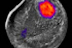
Elastography has been drawing interest as a means to use ultrasound to detect pathology by detecting signs of tissue stiffness. In this study, researchers from the University of Washington in Seattle will discuss their method to use elastography on thyroid nodules.Thyroid nodules are a common medical problem, but only 5% are malignant. Biopsy is typically conducted with fine-needle aspiration (FNA); some 300,000 thyroid biopsies are performed each year in the U.S., with 70% of these proving benign.
For the past five years, the authors have been working on a thyroid elastography technique using internal compression provided by the carotid artery. Internal compression has a couple of advantages over external compression, they believe: It reduces operator dependency and the strain estimation error can be minimized, making the technique more repeatable and accurate.
The researchers examined the technique in a population of 82 patients with 92 nodules who were referred for FNA biopsy. Elastography was performed by a commercially available ultrasound machine (HI Vision 5500, Hitachi Medical Systems America, Twinsburg, OH).
Rather than assigning tissue scores based on visual inspection, the researchers calculated an elasticity contrast index (ECI) to quantify the difference in stiffness between a nodule and its surrounding normal tissue. A larger ECI value indicates a stiffer nodule and, thus, an increased chance of malignancy.
The researchers found that the 12 malignant nodules in their patient population had a mean ECI of 6.32 ± 2.25, significantly higher than that of the 80 benign nodules, which had a mean ECI of 3.39 ± 1.58 (p = 0.0008). This indicates that malignant nodules are stiffer than benign nodules compared with surrounding normal tissue.
Using a cut-off ECI value of 3.85 to indicate malignancy, all 12 malignant nodules could be detected, leading to a sensitivity of 100%, specificity of 73.8%, positive predictive value of 36.4%, and negative predictive value of 100%.
The paper will be presented by Yongmin Kim, PhD.

![Examples of ultrasound findings and techniques. (A) Images in a 39-year-old male patient with a mass in the left thigh. The mass is heterogeneous on the B-mode US image (compared with the patient in D) and showed increased microvascularity (superb microvascular imaging [SMI]) and shear-wave elastography (SWE) values. Undifferentiated pleomorphic sarcoma was diagnosed at biopsy (with pleomorphic rhabdomyosarcoma in surgical specimen). (B) Images in an 18-year-old male patient with a mass in the left leg. The mass is hypoechoic on the B-mode image, with no other findings suggestive of malignancy. The lesion is in contact with the cortex of the tibia, which is slightly irregular. CT revealed a doubtful anteromedial tibial erosion. The microvascular study demonstrated high vascularization, suggestive of malignancy. Periosteal Ewing sarcoma was diagnosed with both histologic and immunohistochemical confirmation. (C) Images in a 69-year-old female patient with a lump growing on the outside of the left leg. Multiple SWE examinations were performed (please note the high values obtained in the measurements, whereas the color map highlights the stiffness relative to adjacent tissues). SMI showed areas of increased vascularization to target for sampling. Undifferentiated spindle cell sarcoma was diagnosed at biopsy, with residual leiomyosarcoma in the surgical specimen after neoadjuvant therapy. (D) Images in a 56-year-old female patient with a mass in the right thigh. The mass is heterogeneous at both B-mode ultrasound (similar to patient A) and MRI (coronal T2-weighted spectral attenuated inversion recovery [SPAIR]; T1-weighted pre-contrast and postcontrast imaging), which even shows uptake after the administration of paramagnetic contrast material, which is traditionally suggestive of malignancy. Low values at SMI and elastography are suggestive of benignity. Spindle cell lipoma was diagnosed at biopsy, with atypical spindle cell lipomatous tumor in the surgical specimen.](https://img.auntminnie.com/mindful/smg/workspaces/default/uploads/2026/08/images-radiol250278fig2.APCFLSvX6p.jpg?auto=format%2Ccompress&fit=crop&h=100&q=70&w=100)


![Examples of ultrasound findings and techniques. (A) Images in a 39-year-old male patient with a mass in the left thigh. The mass is heterogeneous on the B-mode US image (compared with the patient in D) and showed increased microvascularity (superb microvascular imaging [SMI]) and shear-wave elastography (SWE) values. Undifferentiated pleomorphic sarcoma was diagnosed at biopsy (with pleomorphic rhabdomyosarcoma in surgical specimen). (B) Images in an 18-year-old male patient with a mass in the left leg. The mass is hypoechoic on the B-mode image, with no other findings suggestive of malignancy. The lesion is in contact with the cortex of the tibia, which is slightly irregular. CT revealed a doubtful anteromedial tibial erosion. The microvascular study demonstrated high vascularization, suggestive of malignancy. Periosteal Ewing sarcoma was diagnosed with both histologic and immunohistochemical confirmation. (C) Images in a 69-year-old female patient with a lump growing on the outside of the left leg. Multiple SWE examinations were performed (please note the high values obtained in the measurements, whereas the color map highlights the stiffness relative to adjacent tissues). SMI showed areas of increased vascularization to target for sampling. Undifferentiated spindle cell sarcoma was diagnosed at biopsy, with residual leiomyosarcoma in the surgical specimen after neoadjuvant therapy. (D) Images in a 56-year-old female patient with a mass in the right thigh. The mass is heterogeneous at both B-mode ultrasound (similar to patient A) and MRI (coronal T2-weighted spectral attenuated inversion recovery [SPAIR]; T1-weighted pre-contrast and postcontrast imaging), which even shows uptake after the administration of paramagnetic contrast material, which is traditionally suggestive of malignancy. Low values at SMI and elastography are suggestive of benignity. Spindle cell lipoma was diagnosed at biopsy, with atypical spindle cell lipomatous tumor in the surgical specimen.](https://img.auntminnie.com/mindful/smg/workspaces/default/uploads/2026/08/images-radiol250278fig2.APCFLSvX6p.jpg?auto=format%2Ccompress&fit=crop&h=112&q=70&w=112)