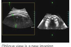
WASHINGTON, DC - Three-dimensional (3D) ultrasound can accurately detect abnormal fetuses, according to a presentation at this week's American Institute of Ultrasound in Medicine (AIUM) annual meeting.
During a categorical course at the AIUM meeting, Dr. Beryl Benacerraf from the Harvard Medical School in Boston presented the results from a study of 26 fetuses.
"All of the anomalous fetuses were correctly identified (using 3D), although not all of the individual anomalies were correctly seen," Benacerraf said.
The Harvard researchers used 2D and 3D ultrasound to evaluate the 26 fetuses, of which six were normal and 20 were abnormal. After a standard 2D sonogram was performed, a sonographer obtained five volume acquisitions designed to encompass the entire fetal anatomy.
Several weeks later, a physician (Benacerraf), without knowledge of the original clinical findings, reviewed the 3D volumes. The normal and abnormal cases were randomized.
Based on the offline review, Benacerraf categorized the studies as normal or abnormal, and compared the results with the original 2D exam findings. For the purposes of the study, the 2D findings were used as the gold standard.
The 20 anomalous fetuses were all correctly identified as abnormal based on the 3D review. On the 2D exams, 57 anomalies were found, 49 (86%) of which were identified on 3D. There were two false-positive findings seen on 3D.
Anomalies missed on 3D included micrognathia (two of five cases missed), polydactyly (two of three cases missed), and one case each of hypertelorism, a small liver cyst, and a small bowel containing an omphalocele. These anomalies occurred in six fetuses with other anomalies that were identified on both 2D and 3D, Benacerraf said.
"While the offline 3D volume has been shown to be accurate in screening for fetal anomalies for the most part, once the fetus is identified as abnormal, then a full 2D examination by a specialist should be performed because obviously we're not picking up all of the details of all of the abnormalities in a single fetus,” she said.
By Erik L. Ridley
AuntMinnie.com staff writer
March 24, 2006
Related Reading
3D imaging creates paradox for ultrasound vendors, November 9, 2005
Practical tips ease 3D/4D ultrasound, August 12, 2005
3D offers speed gains for fetal scanning, July 1, 2005
AMA says ultrasound in-utero 'portraits' are bad idea, June 22, 2005
4D ultrasound shows promise for improving fetal echocardiography, April 6, 2005
Copyright © 2006 AuntMinnie.com